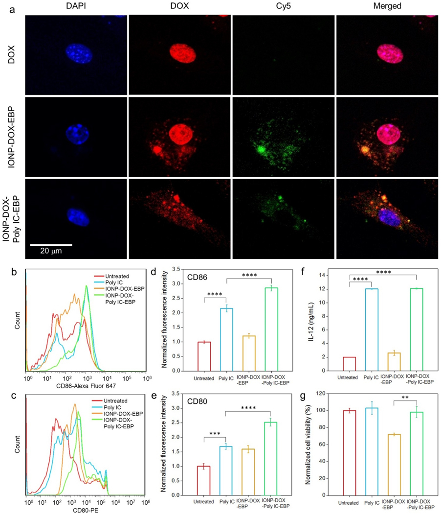
Fig. 3.
Cellular responses of BMDCs to various agents. For all assays, 10 μg/mL Poly IC or an agent carrying equivalent Poly IC was incubated with cells. a, Confocal fluorescence microscopy imaging of cellular uptake of free DOX, IONP-DOX-EBP and IONP-DOX-Poly IC-EBP into BMDCs. Cell nuclei were stained with DAPI. DOX was imaged at Ex: 495 nm and Em: 580–654 nm. NPs were labeled with Cy5 (red) and imaged at Ex: 652 nm and Em: 665–745 nm. b-c, Flow cytometry study of DC maturation. BMDCs were incubated with Poly IC, IONP-DOX-EBP or IONP-DOX-Poly IC-EBP for 24 h and the expression of (b) CD86 and (c) CD80 were evaluated. d-e, Mean fluorescence intensities of anti-CD86 and anti-CD80 antibodies, respectively, derived from (b) and (c). f, Production of IL-12 by BMDCs in cellular supernatants 24 h after incubation with Poly IC, IONP-DOX-EBP or IONP-DOX-Poly IC-EBP, quantified by ELISA. g, Toxicity of Poly IC, IONP-DOX-EBP and IONP-DOX-Poly IC-EBP on BMDCs after 24 h incubation, assessed by the Alamar Blue viability assay. **P < 0.01, ***P < 0.005, ****P < 0.0001 by one-way ANOVA with Turkey’s post-hoc test.

