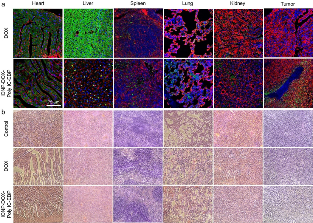
Fig. 7.
Uptake of DOX into tumor and tissue histology of various organs in mice treated with free DOX or IONP-DOX-Poly IC-EBP. BALB/c mice were treated with saline (negative control), DOX (positive control) or IONP-DOX-Poly IC-EBP (therapeutic agent). DOX dose was 10 mg/kg and Poly IC dose was 18 mg/kg, and tissues were collected 48 h after a single i.v. injection. a, Confocal fluorescence microscopy imagines of tissues sections of various organs/tumors. Red: cell membrane (WGA-AF647). Blue: cell nucleus (DAPI). Green: DOX (Ex: 495 nm; Em: 600–650 nm). Scale bar: 75 μm. b, H&E stained tissue sections of heart, liver, spleen, lung, kidney and tumor from mice treated with the same conditions in a.

