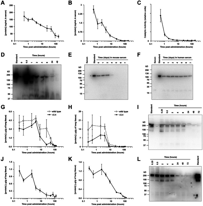
Figure 3. Pharmacokinetics of sACE22.v2.4-IgG1 peptide.
(A-C) sACE22.v2.4-IgG1 was IV administered to mice (N=6 per time point; 2.0 mg/kg). Serum was collected and analyzed by human IgG1 ELISA (A), by ACE2 ELISA (B), and for ACE2 catalytic activity (C). (D) Serum samples from representative male mice were separated on a non-reducing SDS electrophoretic gel and probed with anti-human IgG1 using 10 ng of purified sACE22.v2.4-IgG1 as a standard. Predicted molecular weight (MW; excluding glycans) of dimer is 216 kD. (E-F) sACE22.v2.4-IgG1 was incubated in vitro at 37 °C with normal mouse (E) and human (F) serum. Samples at the indicated time points were separated on a reducing SDS gel and immunoblotted with anti-human ACE2. MW of monomer (excluding glycans) is 108 kD. Shown are representative blots from two experiments. (G-H) Wild type sACE22-IgG1 (white circles) and sACE22.v2.4-IgG1 (black circles) were administered IT at 1.0 mg/kg. Lung tissues were collected, and proteins were extracted and analyzed by (G) human IgG1 ELISA and (H) ACE2 ELISA. N=3 males per time point. (I) Lung extracts from representative mice IT administered sACE22.v2.4-IgG1 were analyzed under non-reducing conditions by anti-human IgG1 immunoblot. (J-K) Mice inhaled nebulized sACE22.v2.4-IgG1. Extracts from lung tissue were analyzed by (J) ACE2 ELISA and (K) human IgG1 ELISA. N=3 males per time point. (L) Representative extracts from lung tissue of mice receiving nebulized sACE22.v2.4-IgG1 were analyzed by anti-human IgG1 immunoblot. Data are presented as mean ± SEM.

