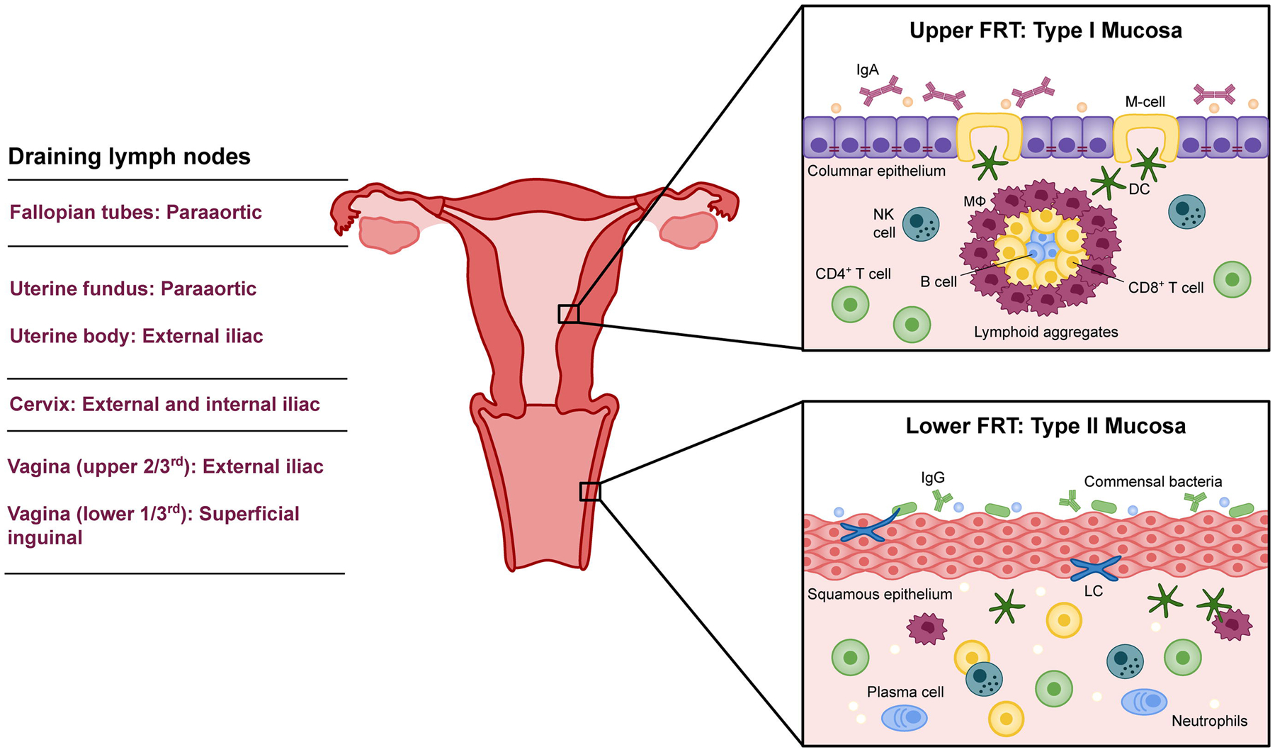
Figure 1. Differences in the immunological milieu of the upper and lower female reproductive tract.

The upper FRT, comprised of the ovaries, fallopian tubes, uterus, and endocervix, is a sterile environment with type I mucosa. As such, it contains organized mucosa-associated lymphoid tissue (MALT) and exerts constitutive immune surveillance via intraepithelial M cells. IgA is the primary immunoglobulin secreted by the upper FRT, though IgG and IgM are also supplied. Antibodies and antimicrobial factors wash into the lower FRT, which is made up of type II mucosa. The ectocervix and vagina do not have MALT or M cells, but possess intraepithelial Langerhans cells which periodically patrol the vaginal lumen. A sequence of draining lymph nodes receive APCs from and prime adaptive immune cells for the FRT.
