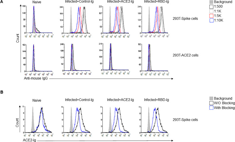
Fig 4. Blocking anti-Spike antibodies are generated in all immunized mice.
(A) Anti-Spike IgG antibodies generated by mice following infection with SARS-CoV-2. At 15 DPI mice were bled through the venous tail and sera were obtained from all mice groups and from naïve mice and diluted as indicated (upper right). Sera was incubated either with 293T-Spike cells (upper histograms) or with 293T-ACE2 cells (lower histograms) as a primary antibody then cells were stained with Alexa fluor 647 anti-mouse IgG secondary antibody. (B) ACE2-Ig staining of 293T-Spike cells in the presence or absence of sera from the various groups. Sera from all indicated groups were incubated with 293T-Spike cells for 1 hour at 4°C followed by staining with ACE2-Ig. All histograms were gated on GFP positive cells. Figure shows one representative experiment out of two performed.

