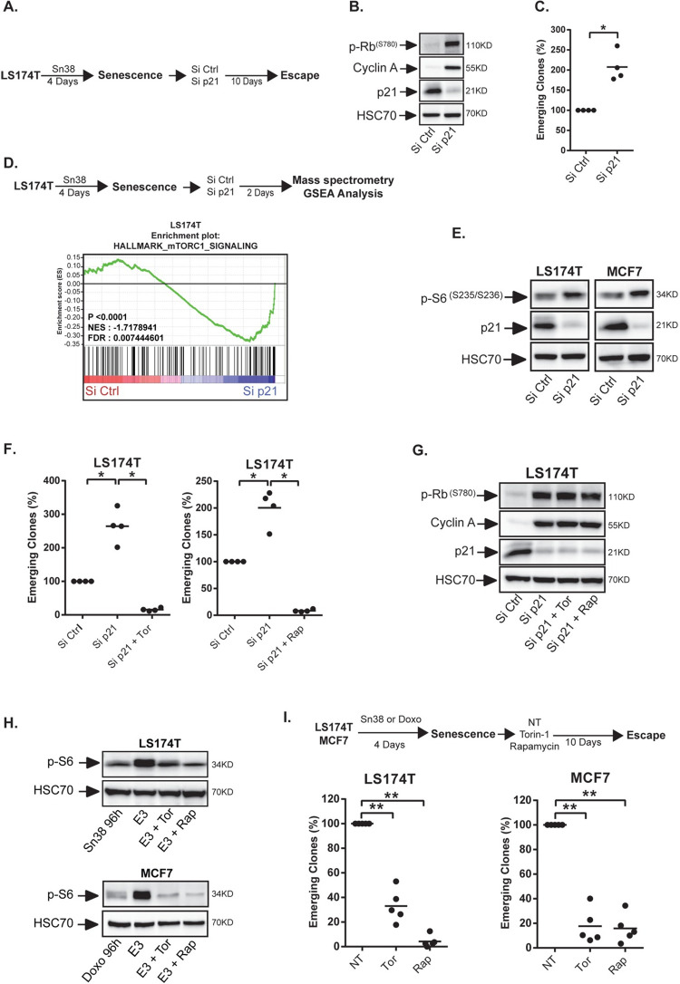
Fig 1. mTOR is necessary for senescence escape.
A. Senescence was induced by treating LS174T colorectal cells with the topoisomerase I inhibitor sn38 as indicated. Cells were then washed with PBS and transfected with a control siRNA or a siRNA directed against p21 for 24 hr and senescence escape was generated by adding 10% FBS. B. LS174T cells were treated as above and cell extracts were recovered 2 days after p21 depletion. The expression of the indicated proteins was analyzed by western blot (n = 3). C. Number of emerging clones analyzed after p21 inactivation (n = 4, Kolmogorov-Smirnov test * = p<0.05). D. Senescent cells were transfected with a control siRNA or a siRNA directed against p21. Cell extracts were analyzed by SWATH quantitative proteomics and GSEA analysis 2 days after p21 depletion (n = 3). E. Validation of mTORCI activation by western blot 2 days after p21 inactivation in LS174T and MCF7 mammary cells (n = 3). F and G. Senescent LS174T cells were transfected with a control siRNA or a siRNA directed against p21 for 24 hr. Cells were then stimulated with 10% FBS in the presence or absence of mTOR inhibitors (Rapamycin: 5nM, Torin-1: 15nM). The number of emerging clones was evaluated after 10 days (F, n = 4, Kolmogorov-Smirnov test * = p<0.05). Cell extracts were recovered after 2 days and the expression of the indicated proteins was analyzed by western blot (G, n = 3). H. Senescent cells were generated as above and emergence was induced by adding 10% FBS. mTORCI activation was analyzed by western blot 3 days after the addition of serum on senescent cells (noted E3 on the figure, n = 3 for MCF7, 2 for LS174T). I. Following senescence induction, cells were stimulated with 10% FBS in the presence or absence of mTOR inhibitors. The number of emerging clones was evaluated 10 days later (n = 5, Kolmogorov-Smirnov test ** = p<0.01).

