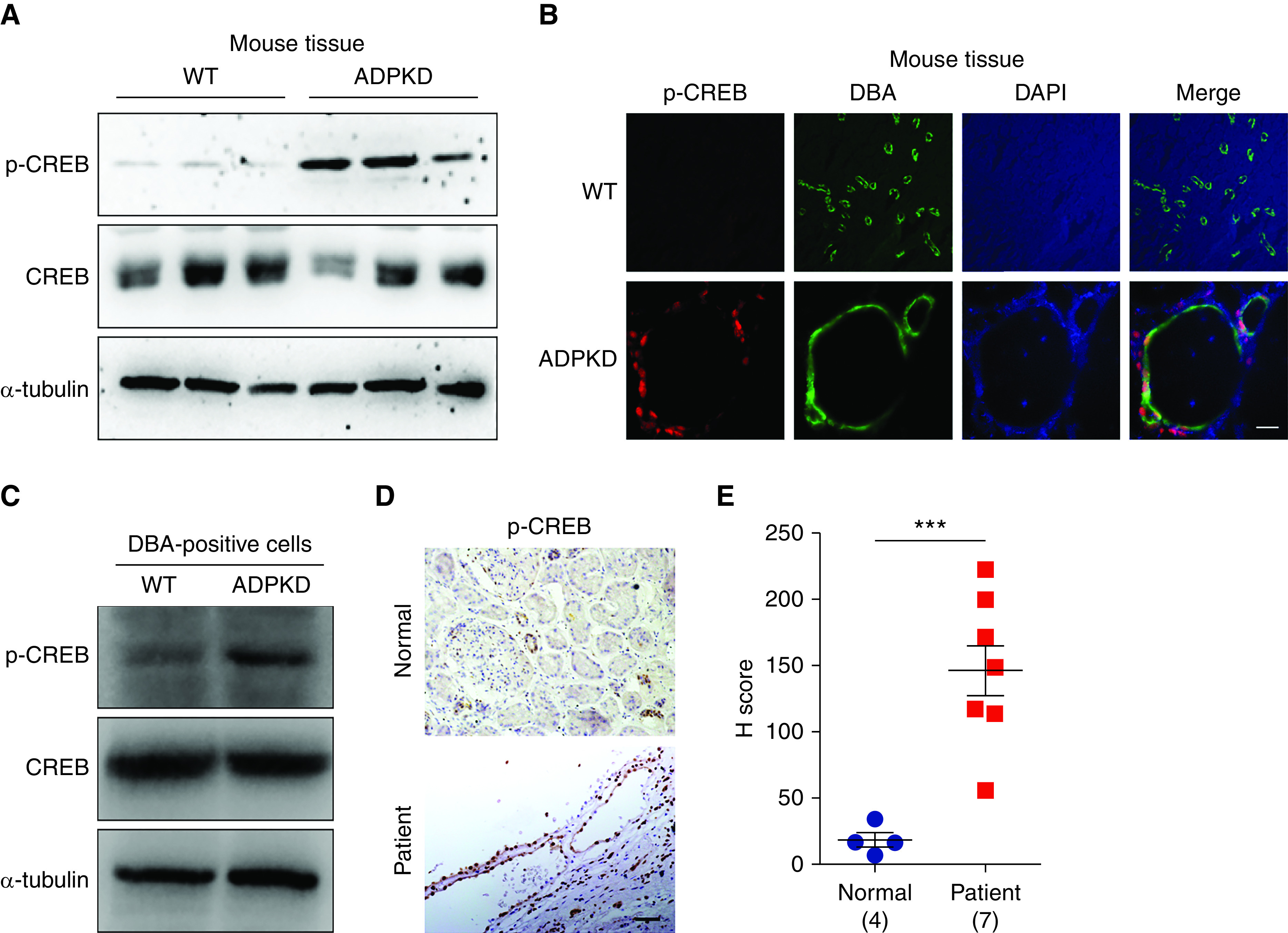
Figure 1.

Phosphorylation of CREB is increased in ADPKD kidneys. (A) Western blot analysis of p-CREB (Ser133) in kidneys from WT and early-onset ADPKD mice. (B) Immunofluorescence analysis of p-CREB level in kidneys from WT and ADPKD mice. Scale bar, 25 μm. Original magnification, 650×. (C) Western blot analysis of p-CREB in DBA-positive cells from WT and ADPKD mice. (D) Immunohistochemistry analysis of p-CREB level in normal kidney and kidney from a patient with ADPKD. Scale bar, 50 μm. Original magnification, 200×. (E) The signal density of p-CREB was determined by the H score. Data are represented as means±SEMs, and were analyzed using the unpaired, two-sided t test. ***P<0.001. DAPI, 4′,6-diamidino-2-phenylindole.
