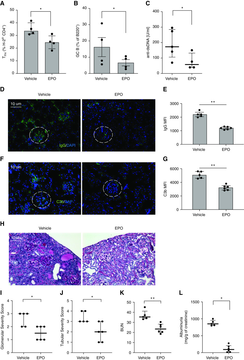
Figure 8.
EPO reduces TFH cell induction, GC B cells formation, and anti-dsDNA formation in the B6 into B6 x DBA parent to F1 model of lupus. B6 spleen cells were injected into B6xDBA (bxd) recipients that were treated with EPO (10,000 IU/kg/day, i.p.) or vehicle control until euthanasia on week 10 after injection. Percentages of TFH (A) and GC B cells (B) at sacrifice. Anti-dsDNA autoantibodies in sera collected from the same mice at sacrifice (C). Representative images and quantification of IgG (D), (E) and C3b (F), (G) glomerular deposition in staining of kidney tissues. Original magnification ×20. Differences in IgG (G) and C3b (I) glomerular fluorescent intensity between EPO- and vehicle-treated animals were quantified relative to DAPI using MetaMorph software. At least 20 glomeruli from three animals were included in the analysis. Scale bars: 10 μm. Representative images of periodic acid–Schiff staining of kidney tissue (H). Quantification of glomerular (I) and tubule-interstitial (J) scores, and BUN (K). Albuminuria (L) expressed as the ratio of urine albumin to creatinine. Data represent median±IQR. *P<0.05; **P<0.01. Data were analyzed using unpaired Mann–Whitney test.

