Figure 4.
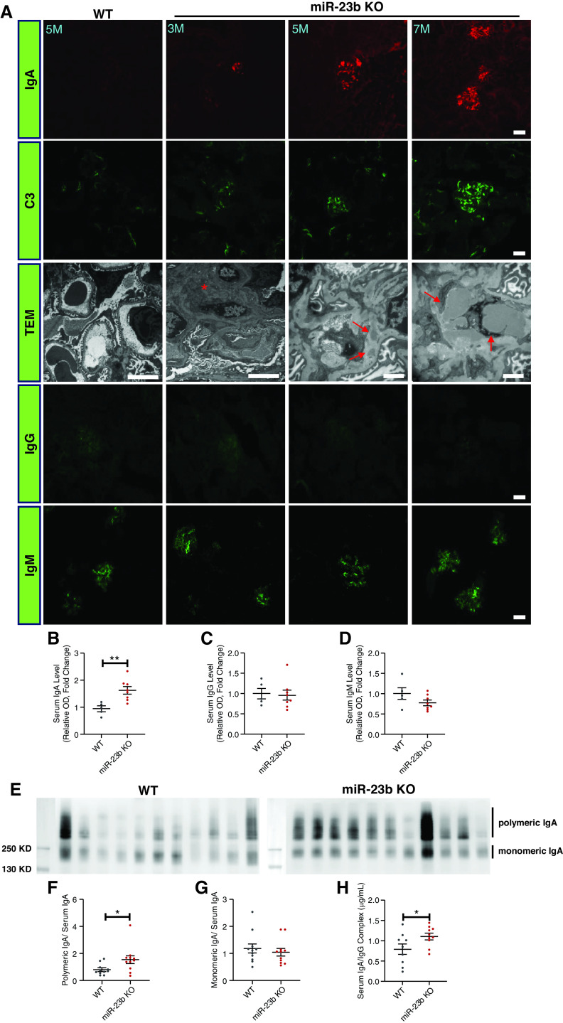
miR-23b−/− mice develop an IgAN-like disease. (A) Representative images of IgA, C3, IgG, and IgM immunofluorescence staining of kidney sections and TEM. TEM images demonstrate the presence of electron-dense mesangial deposits (red arrows) consistent with mesangial IgA immune complex deposition in miR-23b−/− mice. Quantification of (B) serum IgA, (C) IgG, and (D) IgM levels in 5-month-old WT (n=5) and miR-23b−/− (n=8) mice. SDS-PAGE and IgA western blotting of serum IgA from (E) 5-month-old WT (n=11) and miR-23b−/− (n=11) mice with (F) densitometric analysis of polymeric IgA: total serum IgA and monomeric IgA: (G) total serum IgA. (H) Levels of IgA-IgG immune complexes in the sera of 5-month-old WT (n=9) and miR-23b−/− (n=10) mice.

