Figure 6.

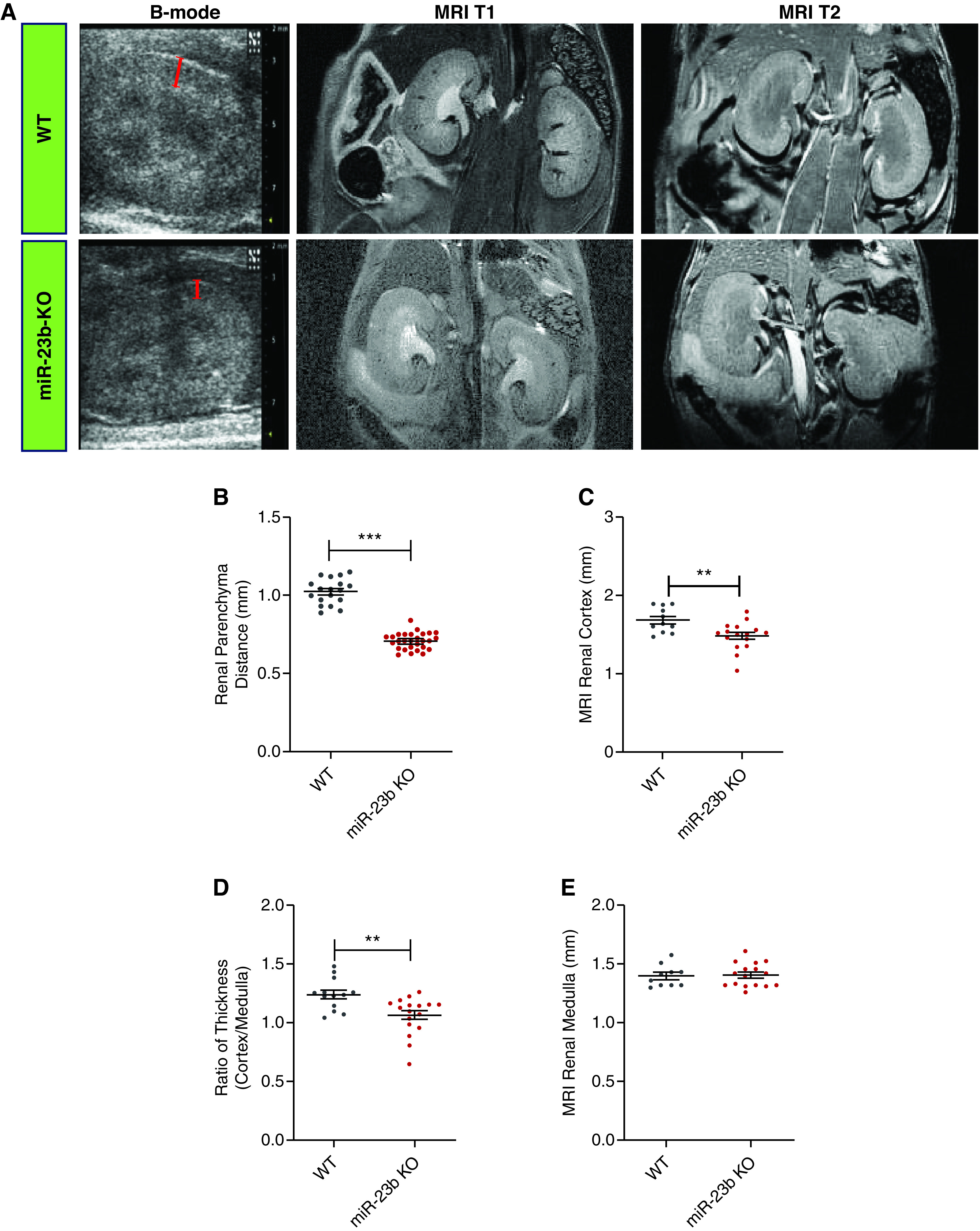
Deletion of miR-23b induces structural kidney changes. (A) Representative images of B-mode ultrasonography of the kidney (first panel, bars estimating the renal parenchyma distance) in WT (n=5) and miR-23b−/− (n=7); MRI T1/2 images (second and third panels) in WT (n=10), and miR-23b−/− (n=12) 5-month-old male mice. (B) Quantification of the renal parenchymal thickness measured from kidney ultrasound images. (C) Quantification of the renal cortical thickness measured from MRI T2 images. (D) Quantification of the ratio of MRI T2 cortex to medulla thickness. (E) Quantification of MRI T2 medulla thickness; *P<0.05, **P<0.01, and ***P<0.001. Data are shown as the mean±SEM.
