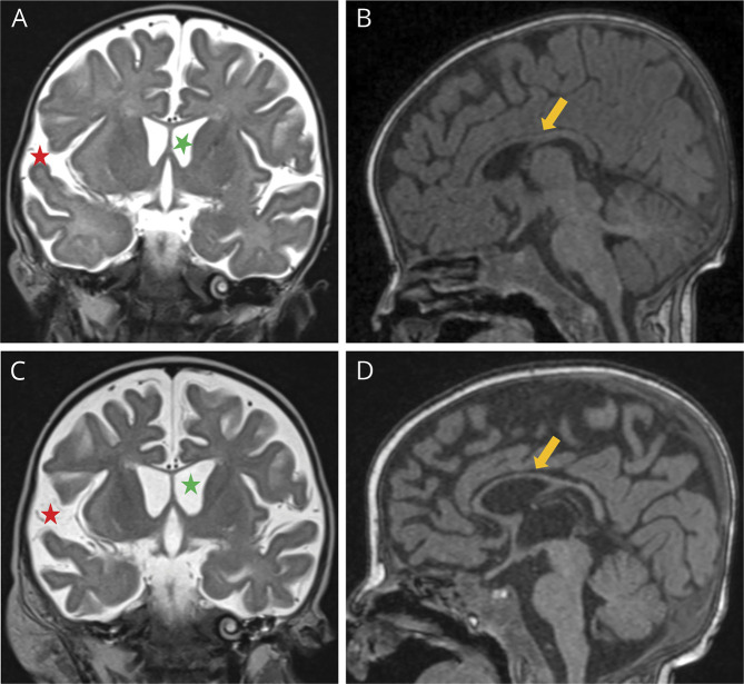
Figure 1. Brain MRI at 3 and 4 Months of Life, Respectively.
In the first row, images of the first MRI study (A and B), performed before liver transplantation, demonstrated enlarged ventricular system and subarachnoid spaces (green and red asterisks, respectively, in A) and reduced thickness of the corpus callosum (yellow arrow in B). In second MRI study in the second row (C and D), performed 2 weeks after liver transplantation, a progression of these findings was noted (asterisks in C and arrow in D).

