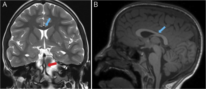
Figure 2. Brain MRI at 9 Years of Age.
Last MRI study (A and B) was performed 9 years later, after normalization of growing curves, and demonstrated a regression of brain atrophy and normal thickness of the corpus callosum (blue arrows in A and B comparing with yellow arrows in B and D of Figure 1). As an incidental finding, a basilar artery's fenestration (anatomic variant) was seen in (A) (red arrow).

