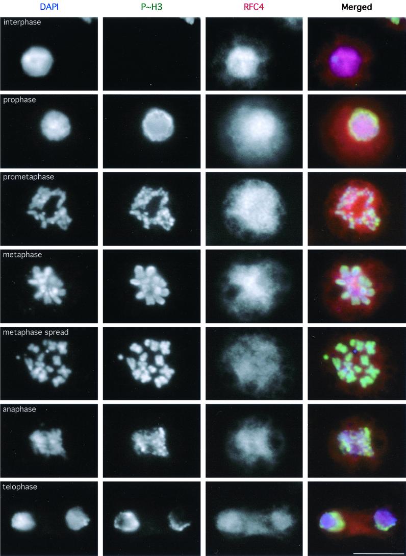
FIG. 5.
Detection of DmRFC4 by immunofluorescence in mitotic Drosophila cultured cells. Dmel2 cells were processed for immunofluorescence detection of RFC4. These cells were also stained for the mitosis-specific phosphorylated form of histone H3 to unambiguously distinguish mitotic cells (18). RFC4 appears to be homogeneously distributed throughout mitotic cells from prophase through telophase. A metaphase cell processed to spread the chromosomes is also shown. Scale bar = 10 μm.

