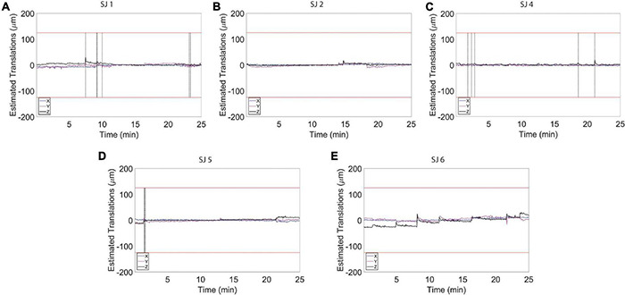
FIGURE 6.

Estimated motion levels for individual animals during auditory functional magnetic resonance imaging (fMRI) acquisition. (A–E) Estimated motion levels (translations) for five individual animals during auditory fMRI acquisition. Motion levels during the auditory task are lower and more stable compared to the levels detected during the resting state acquisitions (Figure 4). The red lines represent 1/4 of the voxel size. The vertical black line indicates the location of a spike in the time course that was removed using the Matlab toolbox, ArtRepair.
