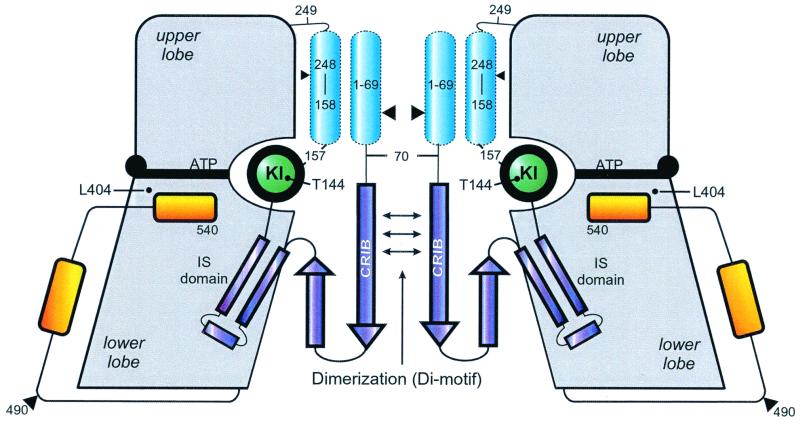
FIG. 1.
Structural model of αPAK, using as a guideline the X-ray structure of a complex between a regulatory fragment from residues 70 to 175 and the complete CD (residues 249 to 545) from human αPAK (18). The regulatory part of the molecule is in dark blue structural elements not included in the three-dimensional structure are in light blue. Approximate locations of residues relevant to the results presented in this report are indicated. It was previously assumed that inhibition by the inhibitory switch (IS) domain and dimerization via the Di motif is relieved by binding of Cdc42 to the CRIB region. KI, kinase inhibitory linker.

