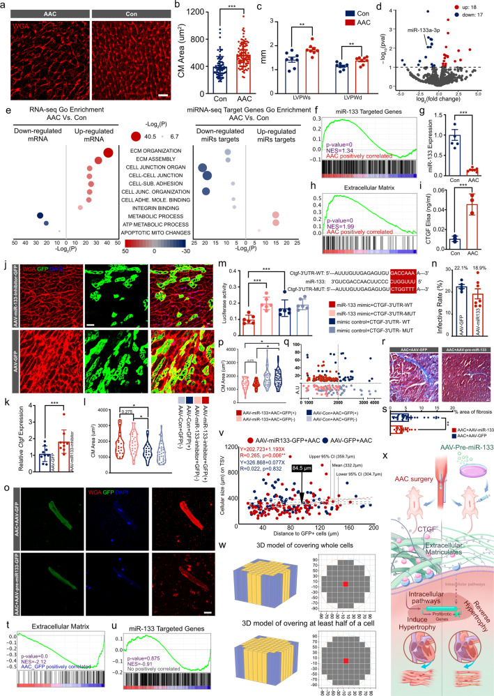
Fig. 1.
Exceeding mechanical stress induced CM hypertrophy via miR-133-CTGF-ECM-integrin-Hippo-YAP mechanotransduction signaling. Targeting ECM remodeling through miRs based therapy attenuated hypertrophic phenotype, and AAV strategy delivering miR-133 could prevent pressure overload-induced hypertrophy both in infected and surrounding non-infected CMs, which indicated a modified AAV administration approach with a reduced dosage down to a theoretic calculating value as 1.6% of infected rate to achieve therapeutic propose. a, b The WGA staining results of heart tissues (N = 5 biologically independent samples), and the cell size was measured in 10 fields/slice in both groups (n > 20 CMs for each individual animal). c The echocardiographic results of AAC mice demonstrated the thickness of LVPW both in systolic and diastolic period. d Volcano plot showing the log fold changes and log p-value of each gene. Up-regulated genes were marked red and down-regulated genes were marked as blue. And miR-133a-3p ranked one of the most differentiated genes. e Scatter plot displaying the differentiated Go term enrichments in AAC hearts of RNA-seq and differentiated miRs targeting genes of miR-seq. f GSEA with the genes as miR-133 targets revealed up-regulated enriched in AAV hearts. h Another GSEA indicted the gene ontology gene-sets of ECM-related pathway up-regulated enriched in AAC hearts. g qPCR demonstrated the inhibition of miR-133 in AAC hearts. i CTGF was significantly elevated by Elisa Assay. j–l Hypertrophy of CMs had been recorded both in infected and non-infected cells after AAV-miR-133-inhibitor-GFP administration (N = 5 biologically independent samples, n > 20 CMs for each individual animal). k Cgft expression up-regualted after miR-133 inhibition. m The putative binding sites located in the 3′ UTR of Ctgf and the experimental design of luciferase reporter. H293T cells were co-transfected with Ctgf-WT or Ctgf-MUT and miR-133 or miR-NC. The luciferase activity was analyzed. n Isolated CMs were measured cellular size after AAV-pre-miR-133-GFP administration. o Infected rated were calculated in heart cryo-sections (N = 4 biologically independent samples). p, q Violin and scatter plots showing the cellular area distribution with differentiated GFP density. Results revealed the attenuation of hypertrophy both in GFP+ and GFP− CMs (N = 5 biologically independent samples, n > 20 CMs for each individual animal). r, s Masson staining showing the reduction of fibrotic area after AAV-pre-miR-133 administration (N = 3 biologically independent samples). t GSEA demonstrated the gene ontology gene-sets of ECM-related pathway dysregulation in AAV delivering miR-133 hearts based on RNA-seq. u GSEA showed no positive correlation of miR-133 targeted genes in Yap1-overexpression hearts. v Linear regression to identify the distance of one infected to affect surrounding CMs. And a distance of 84.5 μm was calculated as the efficient distance to reverse hypertrophy among surrounding CMs. w Analysis models had been built to demonstrate the affecting surrounding area with a radius of 84.5 μm, following two kinds of principles as covering whole cells or at least half of a cell, which indicated a minimal infected rate of 1.67% or 0.64% to achieve efficient heart AAV based therapy in theory, respectively. x Graphic abstract was presented. Scale Bar, 50 μm; all the data were shown as mean ± SD, *P < 0.05; **P < 0.01; ***P < 0.001. AAC abdominal aorta contraction, AAV adeno-associated virus, CI confidence interval, CM cardiomyocyte, ECM extracellular matrix, LVPWs left ventricular partial wall, systolic, LVPWd left ventricular partial wall, diastolic, NES normalized enrichment score, WT wild type, Yap1-OE Yap1 overexpression

