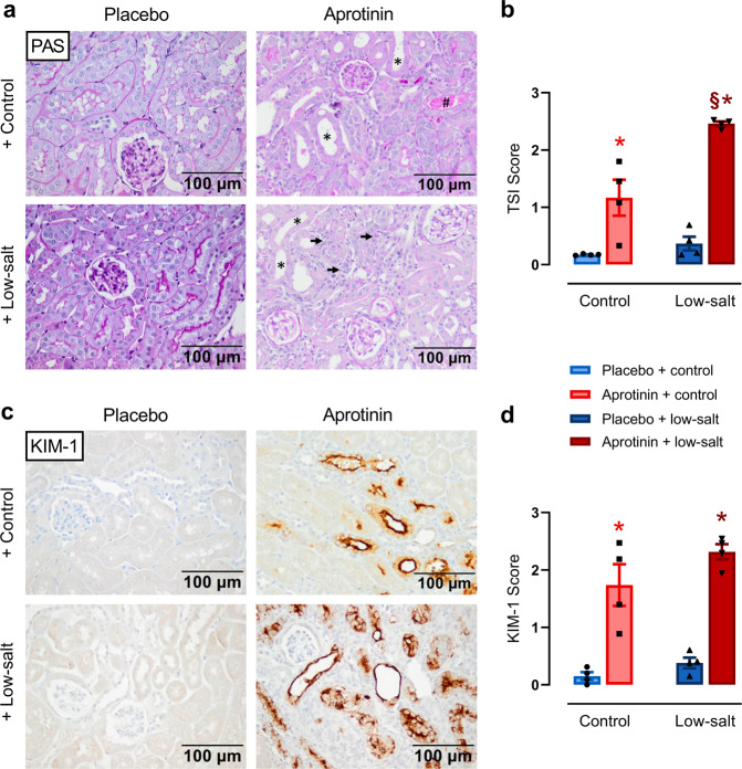
Fig. 5. Effect of aprotinin on kidney histology.
a PAS stainings from mice treated with placebo or aprotinin under a control or low-salt diet. As expected, kidneys from placebo-treated mice did not show any pathological changes. Kidneys from mice treated with 2 mg aprotinin per day showed moderate tubulointerstitial injury indicated by tubular dilatation and atrophy (marked by *) and rare tubular cast formation (marked by #). In contrast, aprotinin-treated mice on a low-sodium diet developed significantly more severe tubulointerstitial damage, mainly due to a marked increase in interstitial cells, presumably inflammatory cells and fibroblasts (marked by arrows). b Semiquantitative analysis of the observed changes using the tubular sclerosis index (TSI). c Immunohistochemical analysis of the expression of KIM-1, which is a marker of damage to the proximal tubule. d Semiquantitative analysis of KIM-1 expression. (a–d each n = 4). Data and statistical analysis: arithmetic means ± SEM. b, d One-way ANOVA followed by the Dunnett’s test. *P < 0.05, aprotinin vs. placebo treatment. §P < 0.05, low-salt vs. control diet.

