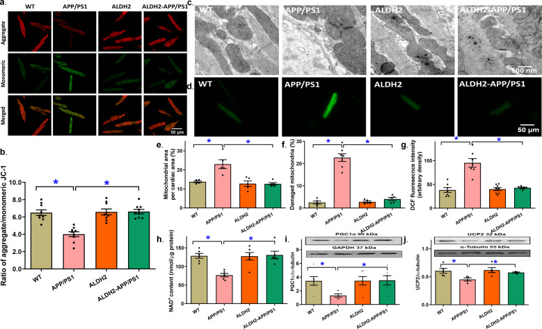
Fig. 4. Mitochondrial membrane potential, mitochondrial permeability transition pore (mPTP) opening, myocardial ultrastructure, and reactive oxygen species (ROS) production in hearts from the APP/PS1 mouse model of Alzheimer’s disease with or without the ALDH2 transgene.
a Representative images of JC-1 fluorescence in cardiomyocytes from various groups (b) Pooled data depicting the ratio of aggregate/monomeric JC-1; (c) Representative transmission electron microscopy (TEM) ultrastructural images (×15,000); (d) Representative DCF fluorescent images depicting ROS production; (e) Mitochondrial area per cardiac area; (f) Percentage of damaged mitochondria; (g) Pooled data from DCF staining depicting ROS levels; (h) mPTP opening evaluated using NAD+ levels; (i) Levels of PGC1α; (j) Levels of UCP2. Insets: representative gel blots showing the levels of the mitochondrial proteins PGC1α and UCP2 using specific antibodies (α-tubulin was used as the loading control). Mean ± SEM, n = 7–9 images or mice per group (n = 5–6 images for panels e, f, and h). *P < 0.05 between the indicated groups.

