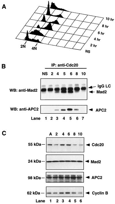
FIG. 1.
Cell cycle-regulated association of Cdc20 with Mad2 and APC. A549 cells were synchronized at the G1/S boundary by a double-thymidine block, released into fresh medium, and collected every hour after release. (A) The profile of DNA content of released cells was analyzed by fluorescence-activated cell sorting. (B) Cells collected at indicated time points (hours) were lysed, cell extracts were immunoprecipitated with goat anti-Cdc20 polyclonal antibody, and the immunoprecipitates were analyzed by Western blotting with mouse anti-Mad2 monoclonal antibody (upper panel) and rabbit anti-APC2 polyclonal antibody (lower panel). Asynchronous cells (lane 1) were included as a control. (C) The levels of Cdc20, Mad2, APC2, and cyclin B proteins at indicated time points (hours) were determined by Western blot analysis. NS, nonsynchronous; IP, immunoprecipitation; WB, Western blotting; IgG, immunoglobulin G; LC, light chain; A, asynchronous.

