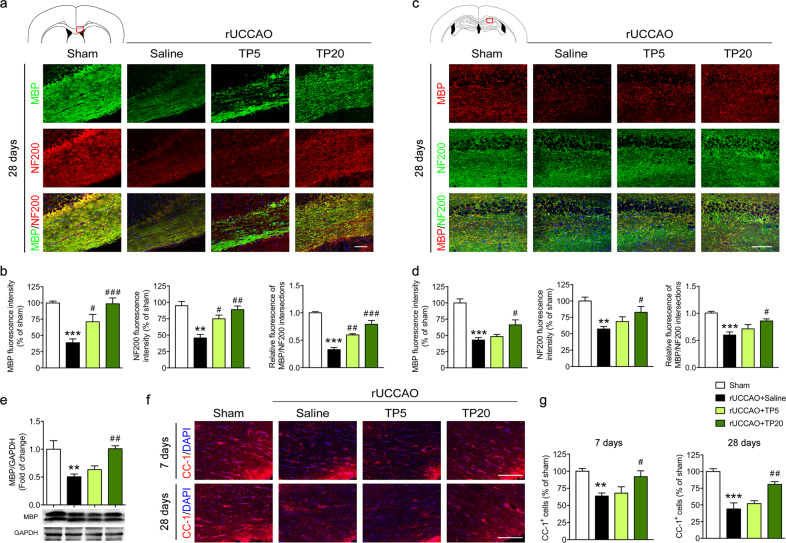
Fig. 1. Triptolide alleviated white matter injury induced by chronic cerebral hypoperfusion.
a, b Immunohistochemical visualization and quantification of MBP and NF200 and colocalization of NF200 and MBP in the corpus callosum (red outlined rectangle in the schematic diagram) at 28 d after right unilateral common carotid artery occlusion (rUCCAO) in mice administered triptolide at a dosage of 5 μg·kg−1·d−1 (TP5) or 20 μg·kg−1·d−1 (TP20). c, d Immunohistochemical visualization and quantification of MBP and NF200 and colocalization of NF200 and MBP in the hippocampus (red outlined rectangle in the schematic diagram) at 28 d after rUCCAO in mice administered TP5 or TP20. e Western blot analysis of MBP expression in the corpus callosum at 28 d after rUCCAO. f, g Immunohistochemical visualization and quantification of CC-1+ oligodendrocyte numbers at 7 d and 28 d after rUCCAO in mice administered TP5 or TP20. Scale bars, 100 μm. n = 3–5 mice per group from at least three independent experiments. **P < 0.01, ***P < 0.001 vs. the sham group; #P < 0.05, ##P < 0.01, ###P < 0.001 vs. the rUCCAO + saline group.

