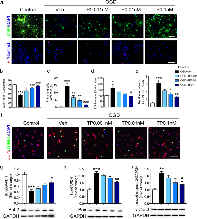
Fig. 3. Triptolide inhibited the apoptosis of oligodendrocytes induced by OGD.
Primary cultured mature oligodendrocytes were subjected to OGD for 2 h and administered with triptolide at concentrations of 0.001, 0.01, and 0.1 nM for 24 h during reperfusion. a, b Immunocytochemical visualization and quantification of the numbers of MBP + mature oligodendrocytes after OGD/reperfusion. a, c Cell death was determined by double-staining with Hoechst 33342 (blue) and propidium iodide (PI, red) in oligodendrocytes after OGD/reperfusion. Scale bars, 50 μm. d Cell damage was evaluated by lactate dehydrogenase (LDH) release assays after OGD/reperfusion. e, f Immunocytochemical visualization and quantification of the numbers of CC-1 and TUNEL double-positive mature oligodendrocytes after OGD/reperfusion. Western blot analysis of the expression of apoptosis-related proteins, including Bcl-2 (g), Bax (h) and cleaved-Caspase-3 (c-Cas3, i), in oligodendrocytes after OGD/reperfusion. n = 3–5 from at least three independent experiments. *P < 0.05, **P < 0.01, ***P < 0.001 vs. the control group; #P < 0.05, ##P < 0.01, ###P < 0.001 vs. the OGD + vehicle (Veh) group.

