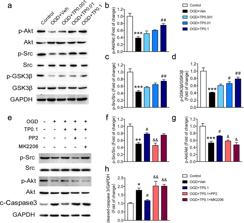
Fig. 4. Triptolide regulated the OGD-induced apoptosis of oligodendrocytes through the Src/Akt/GSK-3β signaling pathway.
a–d Western blot analysis of p-Src, Src, p-Akt, Akt, p-GSK-3β, and GSK-3β expression in oligodendrocytes subjected to OGD for 2 h and administered with triptolide (0.001, 0.01, and 0.1 nM) for 1 h during reperfusion. e–g Western blot analysis of p-Src, Src, p-Akt, and Akt expression in oligodendrocytes subjected to OGD and treated with the Src inhibitor PP2 or the Akt inhibitor MK2206 followed by triptolide (0.1 nM). e, h Western blot analysis of cleaved-Caspase-3 (c-Caspase-3) expression in oligodendrocytes subjected to OGD and treated with the Src inhibitor PP2 or the Akt inhibitor MK2206 followed by triptolide (0.1 nM). n = 3–4 from at least three independent experiments. *P < 0.05, **P < 0.01, ***P < 0.001 vs. the control group; #P < 0.05, ##P < 0.01 vs. the OGD + Veh group; &P < 0.05, &&P < 0.01 vs. the OGD + TP0.1 group.

