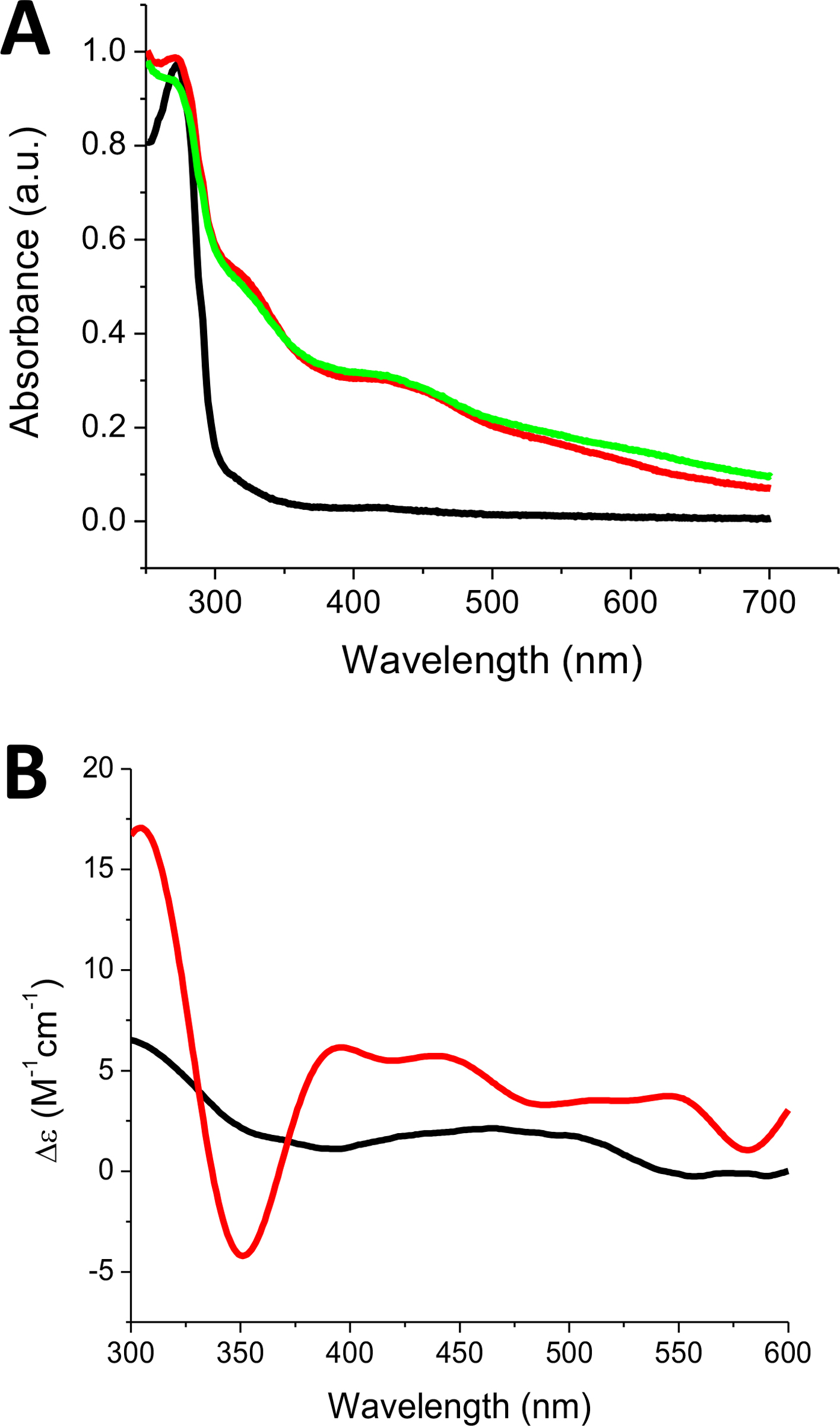
Fig. 5.

Reconstituted holo SyNfu spectra recorded on (A) UV-Vis and (B) CD. Apo SyNfu is shown in (black). For (A), Holo SyNfu reconstituted with Na2S (green) and holo SyNfu reconstituted with Tm NifS and L-Cys (red). For (B), a representative holo spectrum is shown in (red) and apo spectrum shown in (black).
