
Figure 3. ApEn-NK specifically target suspended TNBC cells.
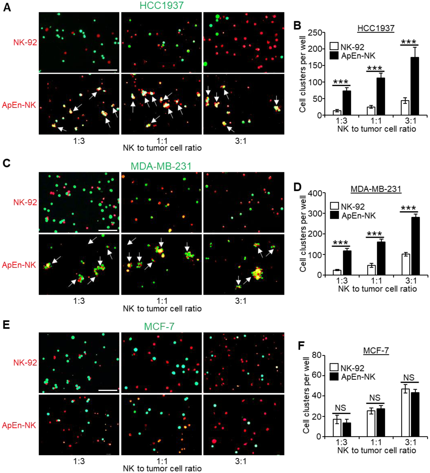
(A-D) ApEn-NK specifically bound and formed more clusters (pointed by arrows) with TNBC cells (HCC1937 and MDA-MB-231) in comparing to baseline controls observed in mixtures with NK92 cells. (E, F) ApEn-NK and parental NK-92 cells showed similar background non-specific binding to non-TNBC cells (MCF-7). Quantitative analyses of cell cluster formation under individual conditions are shown to the right of corresponding microscopic images. Scale bar: 100 μm. All experiments were performed ≥ 3 times with similar results. Representative results are shown. Data are presented as mean ± S.D. ***: P<0.001 (Student’s t-test, two-tailed). NS: not significant.