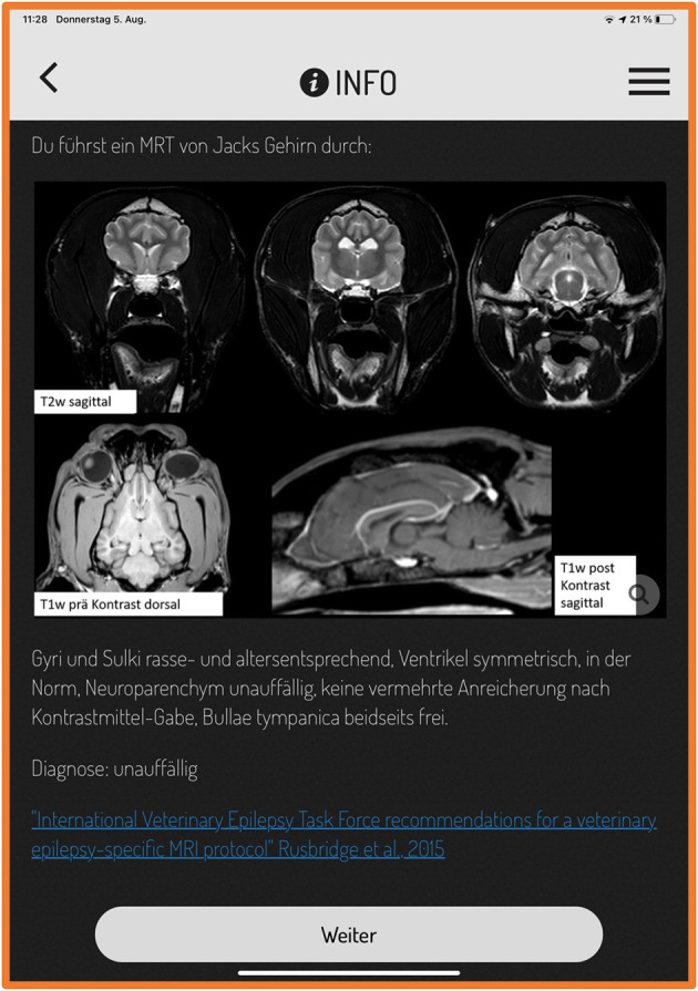
Figure 2.

Original screenshot of actionbound. The screen displays the magnetic resonance imaging (MRI) results of a Labrador Retriever with seizures and a link to an international consensus statement about epilepsy-specific MRI protocols. (Text says: “You performed a MRI of Jack's brain:”; “T2w sagittal”; “T1w pre contrast dorsal”; “T1w post contrast sagittal”, “Gyri and sulci breed and age specific normal. Ventricles symmetrical, normal, brain parenchyma unremarkable, no pathological contrast uptake, tympanic bulla both sides filled with air.”; “next”). T2w, T2 weighted; T1w, T1 weighted.
