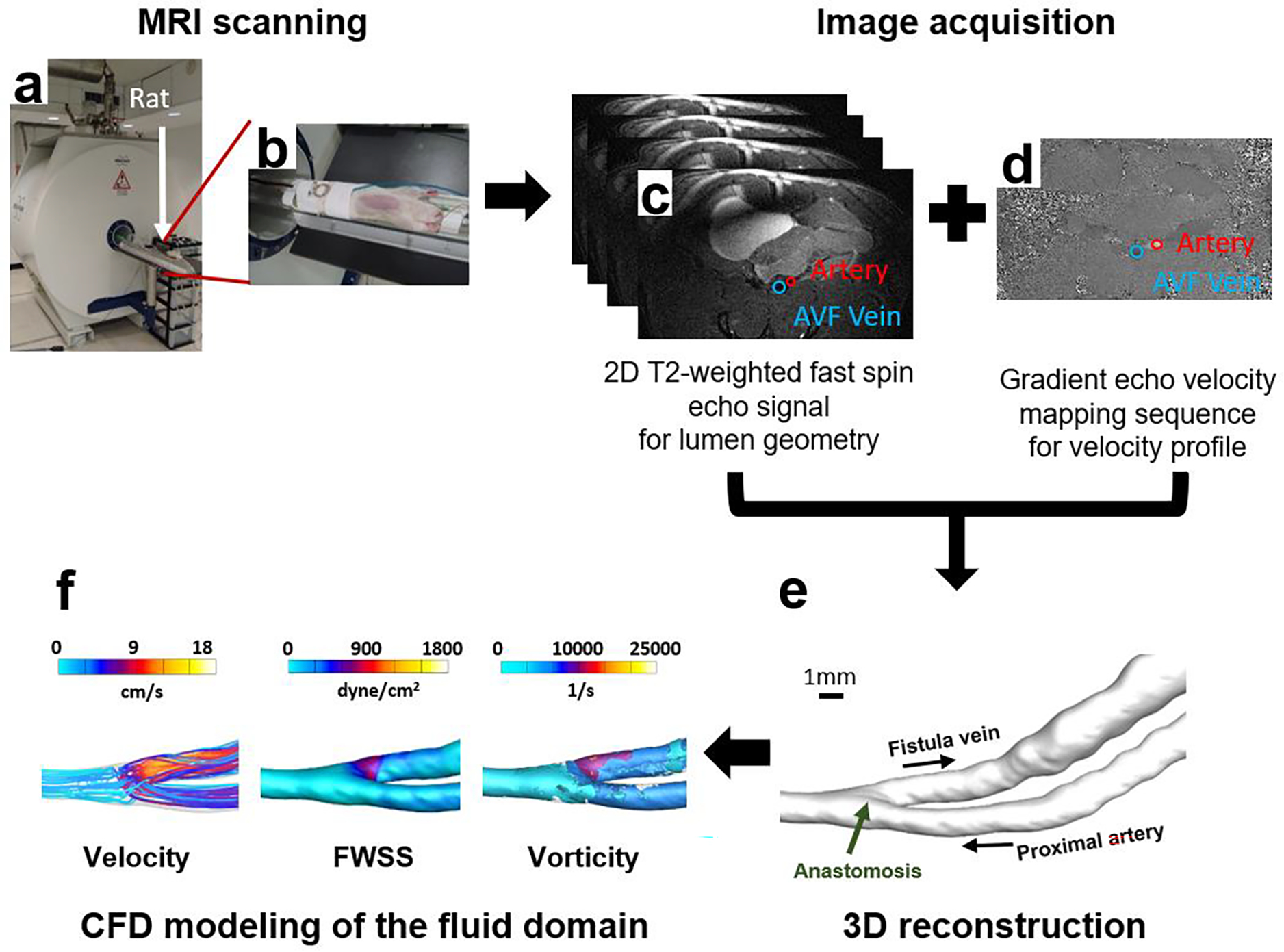
Fig. 6. MRI analysis of rat AVF.

(a), (b) MRI scanning of a rat using a Bruker Biospec 9.4T/20-cm horizontal bore instrument. (c) Image acquisition with a T2-weighted fast spin sequence (black-blood double inversion) for visualization of AVF lumen geometry. (d) Gradient echo velocity mapping sequence for quantitative measures of the blood flow. (e) 3D smoothed geometric reconstruction of the AVF lumen. (f) MRI data extraction and CFD simulation of the fluid domain.
