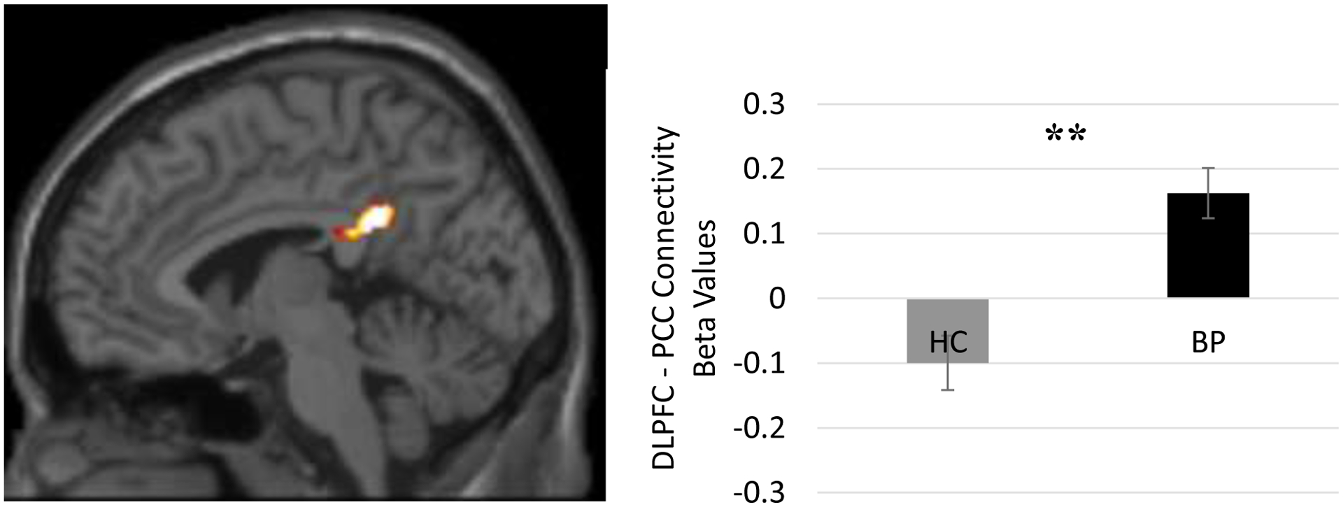
Figure 1. Individuals with BP Pre Treatment > HC Resting State DMN-TPN Functional Connectivity.

Before treatment, there was a significant difference between the bipolar (BP) and healthy control (HC) group in resting state functional connectivity between core regions of the Task Positive Network (dorsolateral prefrontal cortex [DLPFC]) and the Default Mode Network (posterior cingulate cortex [PCC]; t(42) = 4.604, p < 0.001). Individuals with BP showed positively correlated activity between the DLPFC and the PCC (MNI coordinates = −10, −44, 38, k = 153 voxels, Z-score = 4.31). The HC group, in contrast, showed negatively correlated activity between these regions. Note: **p < 0.001
