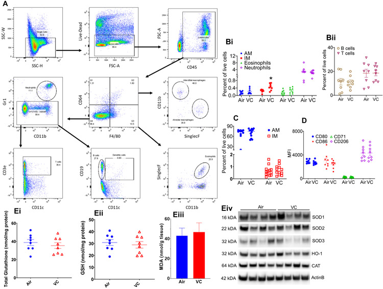
Figure 3: Pulmonary inflammation in vinyl chloride-exposed mice.
(A) Gating scheme for the flow cytometric analysis of immune cells in the lungs and BALF of VC-exposed male C57BL/6 mice. (B) Levels of immune cells – alveolar macrophages, AM; Interstitial macrophages IM; Eosinophils; and Neutrophils (Bi) and B-cells and T-cells (Bii) in the lungs. (C) Abundance of macrophages in the bronchoalveolar lavage fluid (BALF) of VC-exposed male C57BL/6 mice. (D) Analysis of macrophage polarization markers in BALF. (E) Indices of pulmonary oxidative stress: Total glutathione (Ei), GSH (Eii), MDA (Eiii) and western blots of oxidative defense enzymes (Eiv). Values are mean ± SEM. *P < 0.05 vs air-exposed controls.

