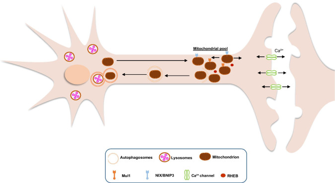
Fig. 2.
Basal neuronal mitophagy. In vivo studies show that PINK1 and Parkin are not activated neither required for mitophagy under physiological conditions. Mitochondria are generated in the soma and transferred in the axons and synapses where there is high ATP and Ca2+ buffering demand. Mitochondria are anchored there but can move anterogradely and retrogradely. PINK1/Parkin-independent mitophagy of depolarized or superfluous mitochondria will initiate until the formation of autophagosomes. Then the autophagosomes will be transported to the soma for fusion with lysosomes and degradation with the help of RHEB. Since PINK1/Parkin signalling is not essential for basal mitophagy, probably Mul1 and NIX/BNIP3 are involved. Mul1, mitochondrial ubiquitin ligase 1; NIX/BNIP3, NIP3-like protein X/BCL2/ adenovirus E1B- interacting protein 3; RHEB, Ras homologue enriched in brain GTP-binding protein

