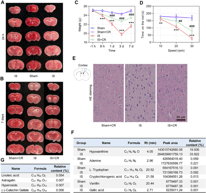
FIGURE 7.
CR treatment significantly ameliorates cerebral ischemic injury. Representative images of TTC-stained brain slices obtained 24 h (A), or 7 days (B) after global cerebral ischemia surgery. (C) Body weight changes were monitored over a 7-day period. (D) The time on the rod in the rotarod test on day 3 (n = 6/group). ** p < 0.01, *** p < 0.001 vs. Sham; ## p < 0.01, ### p < 0.001 IS + CR vs. IS. (E) Representative images of HE staining of the cortex after 7 days of CR treatment. Normal cortical neurons revealed normal cells with rich cytoplasm and round and slightly stained nucleus with relatively large and clear nucleolus formation. Scale bar = 50 µm. (F) The phytochemicals identified by UHPLC/MS/MS in the brains of Sham and IS mice that were orally administered CR. (G) Four components detected only in the brains of CR treated IS mice.

