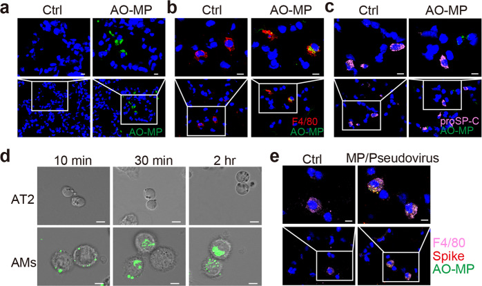
Fig. 2.
Adsorbed virions are delivered into AMs by AO-MPs. ICR mice were intranasally administered PBS (Ctrl) or PKH67-labeled AO-MPs (green, 5 × 10⁶, 50 μl). Thirty minutes later, lung tissues were collected to prepare frozen sections. The AO-MPs in the lung tissues were observed by confocal microscopy (a), and sections were subjected to immunofluorescence staining with anti-F4/80 (red, a marker for macrophages) (b) or anti-proSP-C (pink, a marker for type II cells) (c) antibodies. a-c, Scale bar, 10 μm. d PKH67-labeled AO-MPs (5 × 10⁵) were prepared to adsorb a SARS-CoV-2 pseudovirus at 37 °C for 30 min, and then the virus-adsorbed AO-MPs were incubated with primary AMs and primary alveolar epithelial (AT2) cells isolated from hACE2 transgenic mice for 10 min, 30 min, or 2 h. The uptake of virus-adsorbed AO-MPs by AMs or AT2 cells was analyzed by confocal microscopy. Scale bar, 5 μm. e PKH67-labeled AO-MPs (green) were preincubated with a SARS-CoV-2 pseudovirus for 30 min at 37 °C. Then, the mixture or PBS (Ctrl) was intranasally administered to hACE2 transgenic mice. Lung tissues were subjected to immunofluorescence staining with anti-F4/80 (pink) and anti-spike (red) antibodies. Scale bar, 10 μm

