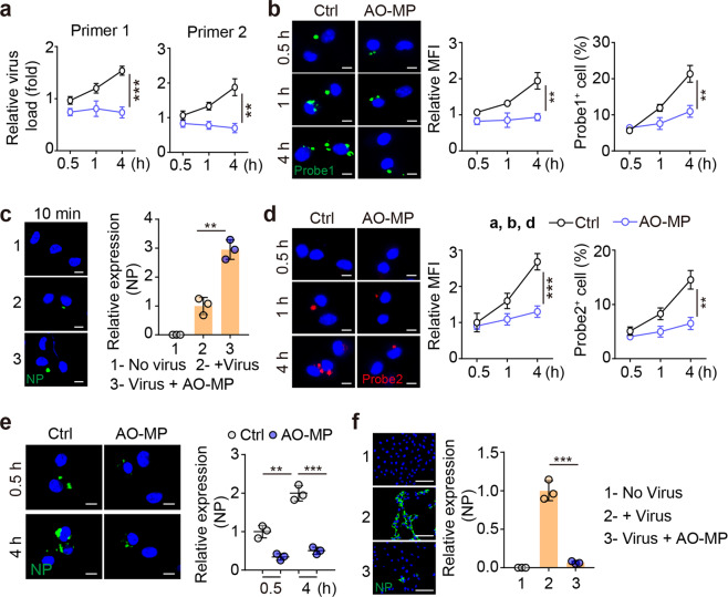
Fig. 3.
AO-MP-delivered SARS-CoV-2 is quenched in AMs. SARS-CoV-2 (5 × 10⁴ TCID50) was incubated with 5 × 10⁵ AO-MPs or PBS (Ctrl) for 30 min at 37 °C and then used to infect AMs. The viral load was determined by real-time PCR with specific primers (a), primer1: ORF1ab gene; primer2: N gene, and cells were fixed for RNAscope analysis (b, d) or immunohistochemical staining for NP (e) at 30 min, 1 h or 4 h post-infection. Probe 1 targeted the viral positive-sense sequence to evaluate the viral distribution (green color) (a), and probe 2 targeted the viral negative-sense sequence to investigate viral replication (red color) (d). Scale bar, 5 μm. c The same as (a), except that cells were fixed for staining with an anti-NP antibody at 10 min post-infection. Scale bar, 5 μm. f The same as (a), except that the supernatants were collected at 4 h to infect Vero E6 cells for 48 h. The cells were stained with an anti-NP antibody. Scale bar, 100 μm. The data represent the mean ± SD of three independent experiments. ** p < 0.01, *** p < 0.001; two tailed Student’s t test (a–f)

