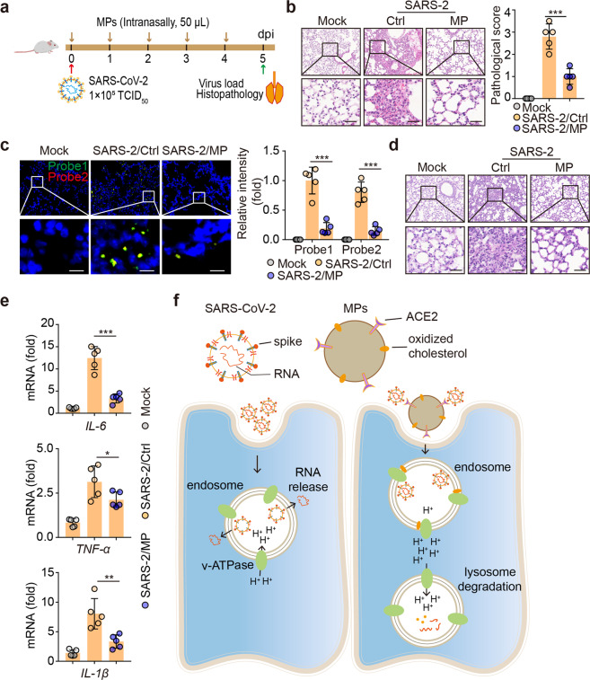
Fig. 7.
Treatment of SARS-CoV-2 infection with AO-MPs in vivo. Schematic diagram of the experimental design. hACE2-transgenic mice were infected with 1 × 10⁵ TCID50 SARS-CoV-2 and then administered AO-MPs (i.n., 50 μl, 5 × 10⁶) once per day for 5 days (a). The control group (Ctrl) received the vehicle (PBS) as a placebo. Lung tissues were fixed for H&E staining (b, n = 5), RNAscope analysis with probes 1 (green) and 2 (red) (c, n = 5), and PAS staining (d, n = 5). Three lung sections from the left lobe were evaluated for each mouse. The representative images selected reflect the distributions of damaged lung tissues. Scale bar, 50 μm for b and d, 10 μm for c. e, The mRNA levels of IL-1β, IL-6 and TNF-α in lung tissues were detected by qPCR. f Schematic of AO-MP-mediated SARS-CoV-2 degradation in AMs. The data represent mean ± SD. * p < 0.05, ** p < 0.01, *** p < 0.001; two-tailed Student’s t-test (b, c) or one-way ANOVA (e)

