Figure 6.

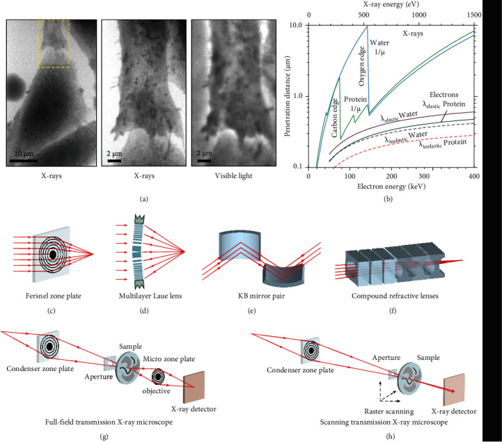
X-ray microscopy setups and their optics. (a) X-ray and optical images of a fibroblast. The area outlined with yellow dashed in the low-magnification image (left panel) is shown at a higher magnification image by the means of X-ray microscopy (middle panel) and optical microscopy (right panel). (b) The penetration distances of X-rays and electrons in water and protein as a function of their energy. The lines from left to right represent attenuation lengths (1/μ) of carbon (protein) and oxygen (H2O) for X-rays and the mean free paths (λ) of H2O (elastic scattering), protein (elastic scattering), H2O (inelastic scattering), and protein (inelastic scattering), respectively. (c) A Fresnel zone plate is made of several transparent and opaque concentric circulars with radially increasing line density. The central absorbing region is responsible for suppressing the strong zero-order diffraction. (d) One-dimensional multilayer Laue lens for hard X-ray focusing. Alternating layers are fabricated by the thin-film deposition technique to implement thin thickness and ultra-high aspect ratio. (e) A Kirkpatrick-Baez mirror for hard X-ray focusing. Multilayer coatings were designed to increase the angles of operation and to perform photon energy selection. (f) Compound refractive lenses for X-ray focusing at a range of 5-40 keV. A linear array of lenses is manufactured by high-density low-atomic number materials. (g) Full-field transmission X-ray microscopy. A full-field image was projected by a microzone plate onto the X-ray detector. (h) Scanning transmission X-ray microscopy. A zone plate is used to focus coherent X-rays on the sample, whereas an X-ray-sensitive detector is used to capture X-ray images. The sample is mounted on a stage having stepping or piezoelectric driven motors to perform the raster scan. (a, b) are reprinted with permission from ref. [88], copyright 1995 Cambridge University Press. (c)–(f) are reprinted with permission from ref. [94], copyright 2010 Macmillan Publishers Limited.
