Fig. 3.
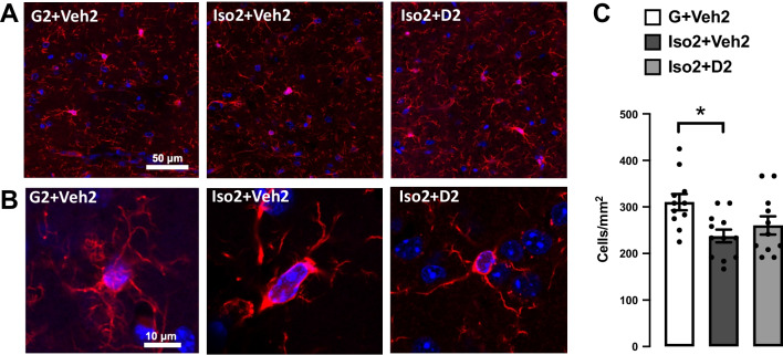
The effects of social isolation-induced anxiety and DHM treatment on microglia activation and proliferation in the hippocampal CA area. A Representative images showing the effect of social isolation-induced anxiety on the number of labeled microglia in the hippocampus, Iba-1 (red), DAPI (blue). B Confocal single-cell microglia images for the G2 + Veh2, Iso + Veh2, and Iso + D2 obtained using a 63X oil-immersion objective. C Quantification analysis of the microglia number in CA1 and CA2 area, data presented as the number of cells per 1 mm2. One-way ANOVA followed by Sidak multiple comparisons test was used for statistical analysis. Each point represents cells number in 2–3 individual sections from n = 5 mice, values represented as mean ± SEM, * = p ≤ 0.05, n = 5

