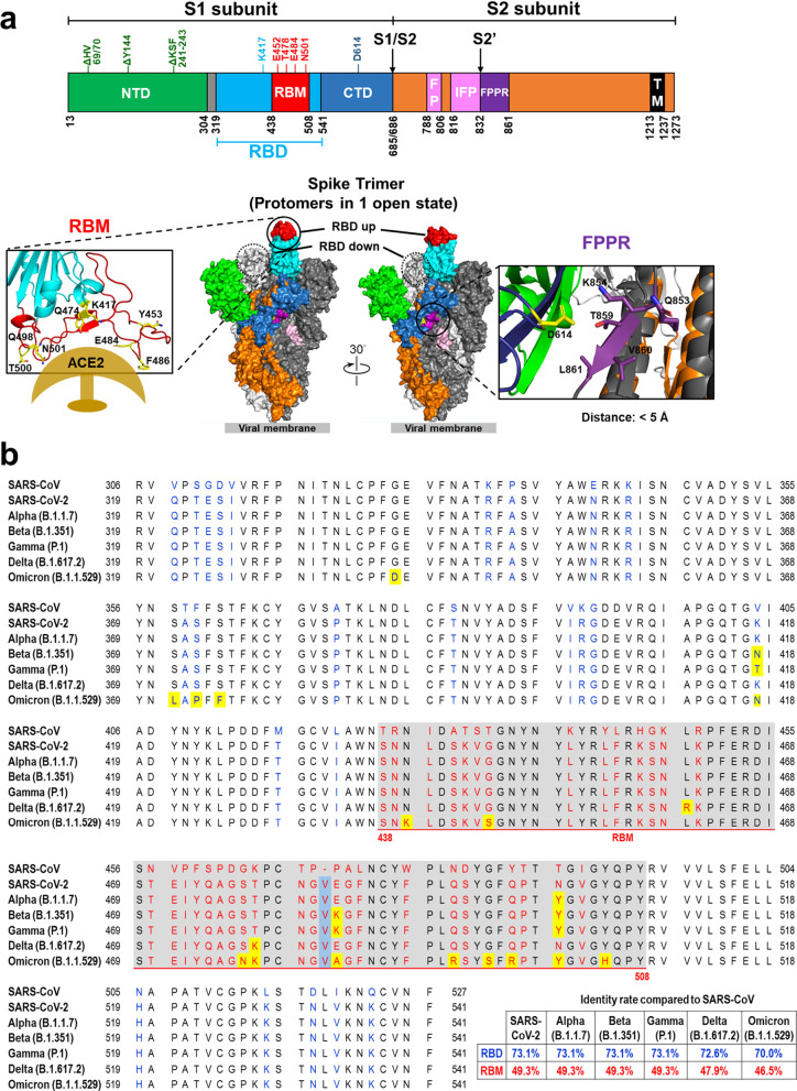
Fig. 1.
SARS-CoV-2 Spike protein. a Structure SARS-CoV-2 spike protein. Different domains of the SARS-CoV-2 spike protein: N-terminal domain (NTD), receptor-binding domain (RBD), receptor-binding motif (RBM), subdomain 1 and 2, protease cleavage sites (S1/S2/S2′), fusion peptide (FP), internal fusion peptide (IFP), fusion peptide proximal region (FPPR), and transmembrane region (TM). HV69/70, Y144, and KSF241-243 are frequently deleted residues in the NTD of SARS-CoV-2 variants of concern. K417, E452, E484, T478, N501 and D614 are the most frequently mutated residues in the RBD of SARS-CoV-2 variants of concern. Key residues of the receptor-binding motif in the S protein of SARS-CoV-2 that interact with ACE2 are shown (lower left). The SARS-CoV-2 S protein trimeric complex is shown in a “one-up” RBD conformation. The two RBD-down protomers are depicted in light and dark gray. The RBD-up protomer is colored according to its domains; RBM in red, non-RBM RBD in light blue, NTD in green, S2 in orange, FP and IFP in pink, and FPPR in purple. The dashed circle indicates the RBD site of an RBD-down conformation protomer. Inter-atomic contacts between aspartate 614 (yellow) in a reference S monomer (dark blue) and five residues (purple) in its adjacent S protein monomer chain (dark gray) within 5 Å. These five contacts might be destabilizing and create a hydrophilic-hydrophobic repulsion that is lost upon replacement of aspartate by glycine in the D614G mutation (lower right). b RBD sequences of SARS-CoV (GeneBank: AAP30030.1), SARS-CoV-2 (GeneBank: QVW76257.1), and SARS-CoV-2 variants of concern, including B.1.1.7 (Alpha), B.1.351 (Beta), P.1 (Gamma), B.1.617.2 (Delta), and B.1.1.529 (Omicron). The amino acids encoded by SARS-CoV-2 that are altered in comparison to SARS-CoV are colored blue (RBD) or red (RBM). The amino acid inserted in SARS-CoV-2 is denoted by a light blue background. The amino acids substituted in variants of concern are denoted by a yellow background. The residues 438–508 comprise the RBM of SARS-CoV-2 and are shown with grey background

