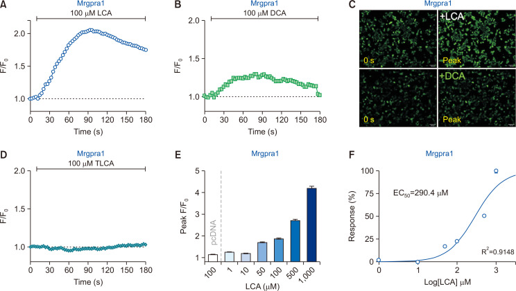
Fig. 1.
Lithocholic acid (LCA) induces an increase in intracellular calcium levels via Mrgpra1. (A, B) HEK293T cells transiently expressing mouse Mrgpra1 (Mrgpra1-HEK293T) were treated with either LCA (100 µM) or deoxycholic acid (DCA; 100 µM) (n=2,932 cells and n=2,312 cells, respectively). (C) Representative fluorescent images of Mrgpra1-HEK293T at initial (0) and peak time points. Scale bar=100 µm. (D) Taurolithocholic acid (TLCA; 100 µM) did not induce any noticeable responses in Mrgpra1-HEK293T (n=3,216 cells). (E) A summary of peak F/F0 from Mrgpra1-HEK293T after treatment with various concentrations of LCA. Cells without Mrgpra1 expression did not show an increase in intracellular calcium levels (pcDNA, n=1,684 cells). (F) Concentration-effect curves with LCA on Mrgpra1-HEK293T (R2=0.9148).

