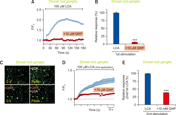
Fig. 5.
Mrgpra1 mediates the effects of lithocholic acid (LCA) in sensory neurons. (A) Mouse dorsal root ganglia (DRG) neurons were treated with LCA (100 µM). An LCA-induced increase in intracellular calcium (n=2,558 cells) was suppressed by pretreatment of 10 µM QWF (n=701 cells) in mouse DRG neurons. (B) A comparison of relative responses between LCA vs. QWF pretreatment. ***p<0.001. (C) Representative fluorescent images of the mouse DRG neurons at initial (0 s) and peak time points for QWF pretreatment before LCA application. Scale bars=100 µm. (D) DRG neurons responded to subsequent LCA exposure (n=155 cells). QWF (10 μM) blocked LCA-induced (100 μM) calcium increase in LCA-responding DRG neurons (n=169 cells). KCl (100 mM) solution was also used to identify viable cells (data not shown). (E) QWF (10 μM) significantly blocked second stimulation of LCA-induced (100 μM) calcium flux in DRG neurons. ***p<0.001.

