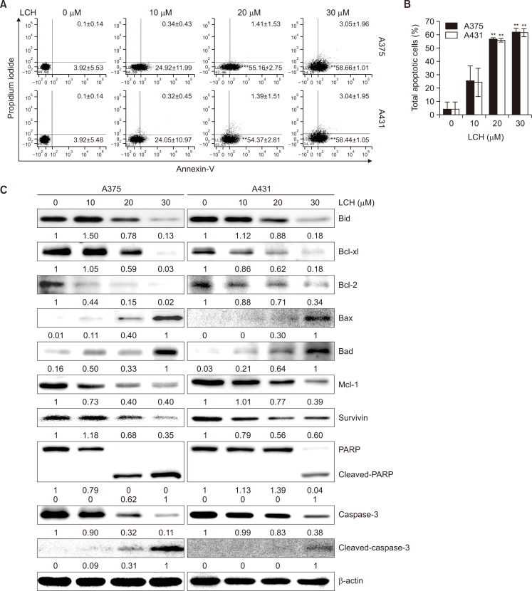
Fig. 2.
Effect of LCH on apoptosis induction in human skin cancer cells. (A, B) Quantitative detection of annexin-V-FITC and PI-positive cells via FACS analysis. A375 and A431 cells were treated with 0, 10, 20, and 30 μM of LCH, and apoptosis was analyzed after annexin-V-FITC and PI double staining. The data represent mean percentages ± SD (n=3; **p<0.01 compared to the control group). (C) A375 and A431 cells treated with 0, 10, 20, and 30 μM LCH for 48 h were harvested to prepare cell extracts. The cell extracts were subjected to SDS-PAGE and Western blot analysis to detect the levels of Bid, Bcl-xl, Bcl-2, Bax, Bad, Mcl-1, survivin, PARP, and caspase-3. β-Actin was used as the denominator to quantify relative protein expression levels.

