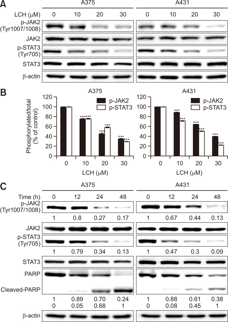
Fig. 4.
Effects of LCH on the modulation of JAK2/STAT3 signaling in human skin cancer cells. (A) A375 and A431 cells were treated with LCH (0, 10, 20, and 30 μM) for 48 h. Whole cell extracts were prepared, separated by SDS-PAGE, and subjected to Western blots using JAK2, p-JAK2, STAT3, and p-STAT3 antibodies. β-Actin was used as the loading control. (B) The percentage of p-JAK2 and p-STAT3 expression is presented. The data represent mean percentages ± SD (n=3; ***p<0.001). (C) Time-dependent effects of LCH on p-JAK2, p-STAT3, and cleaved-PARP were observed using A375 and A431 cells treated with LCH (30 μM) for 0, 12, 24, and 48 h.

