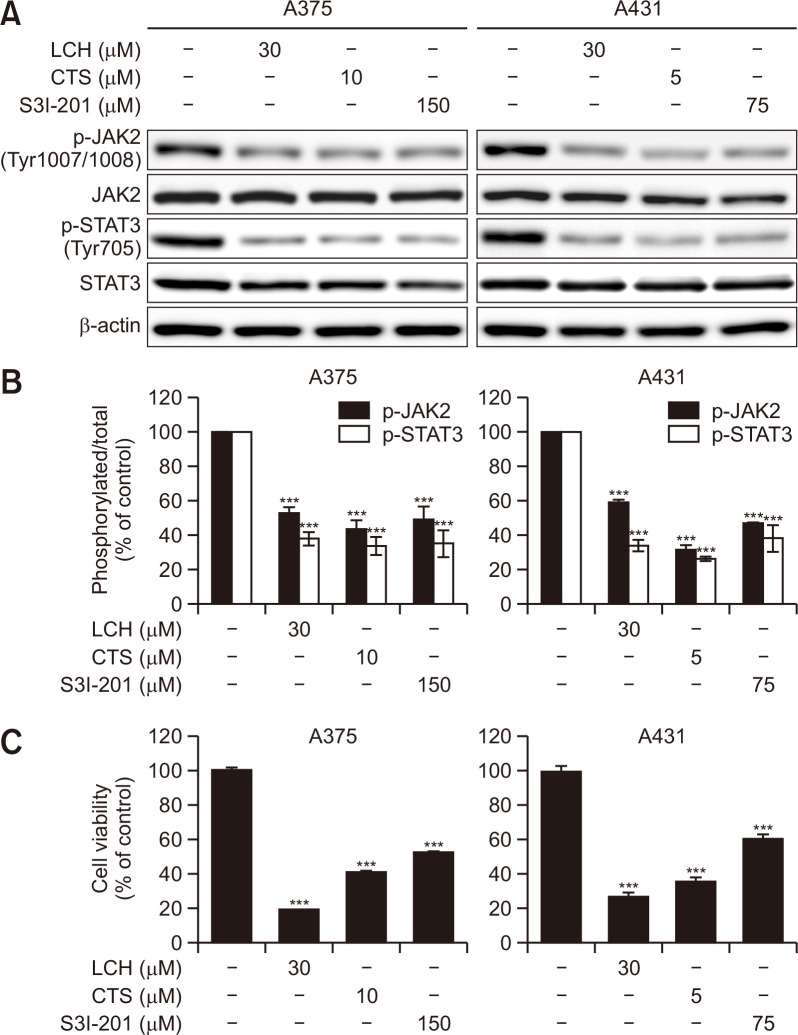
Fig. 5.
Effects of inhibitors of JAK2/STAT3 signaling in human skin cancer cells. (A) A375 and A431 cells were treated with the indicated concentrations of LCH, cryptotanshinone (CTS), and S3I-201 for 48 h. Western blot analysis to detect the levels of JAK2, p-JAK2, STAT3, and p-STAT3. β-Actin was used as the loading control. (B) The percentages of p-JAK2 and p-STAT3 expression is shown. The data represent mean percentages ± SD (n=3; ***p<0.001). (C) Viability of A375 and A431 skin cancer cell lines treated with the indicated concentrations of LCH, CTS, and S3I-201 for 48 h measured using the MTS reagent. The data represent mean percentages ± SD (n=3; ***p<0.001).

