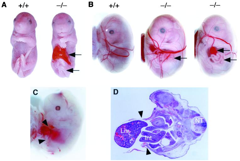
FIG. 4.
Characterization of gastroschisis in ACLP−/− embryos. (A) Wild-type (18.5 dpc) embryo (left) and ACLP−/− sibling (right) exhibiting a disruption of the anterior abdominal wall with herniation of the intestine (upper arrow) and bent tail (lower arrow). (B) Wild-type 15.5-dpc embryo contained within yolk sac (left) and two examples of ACLP−/− embryos with gastroschisis (center and right). ACLP−/− embryo 15.5 dpc with intestine and colon (center, arrow) projecting through a hole in the body wall into the space created by yolk sac, while another ACLP−/− embryo (right) has extruded liver (upper arrow) and intestine (lower arrow). (C) Gastroschisis in 13.5-dpc ACLP−/− embryo. Arrowheads indicate hole with protruding liver. (D) H&E staining of 13.5-dpc ACLP−/− embryo. Arrowheads indicate anterior portion of body wall. Liver (liv), intestine (Int), and neural tube (NT) are indicated.

