FIG. 5.
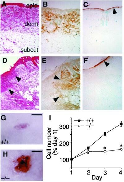
ACLP expression in the skin and defective wound healing in ACLP−/− mice. (A to C) Skin from an uninjured wild-type mouse. (A) H&E staining of skin defining epidermis (epid.), dermis (derm.), and subcutaneous fat (subcut.). Original magnification in panels A to F, ×100. (B) ACLP expression in the dermis. (C) Differentiated epidermal keratinocytes identified by filaggrin immunostaining (arrow). ACLP expression during dermal wound healing. (D to F) Dorsal skin 4 days following a 3-mm punch biopsy. (D) H&E staining showing proliferative keratinocytes (arrow). (E) ACLP expression is confined to the uninjured dermis (arrowheads) and is not expressed in the proliferating keratinocytes. (F) Filaggrin immunostaining (arrowhead). Wild-type and ACLP−/− mice were subjected to a 3-mm punch biopsy through the dorsal skin. Representative dermal wounds from ACLP+/+ (G) and ACLP−/− (H) mice 6 days following punch biopsy (bar = 1 mm). (I) Proliferation deficiency in dermal fibroblasts derived from ACLP−/− mice. Dermal fibroblasts were isolated from 18.5-dpc ACLP+/+ and ACLP−/− embryos and plated in 12-well dishes in triplicate. Cells were counted at 24-h intervals and normalized to cell number at 24 h after plating. ∗, P < 0.05 versus ACLP+/+ control.

