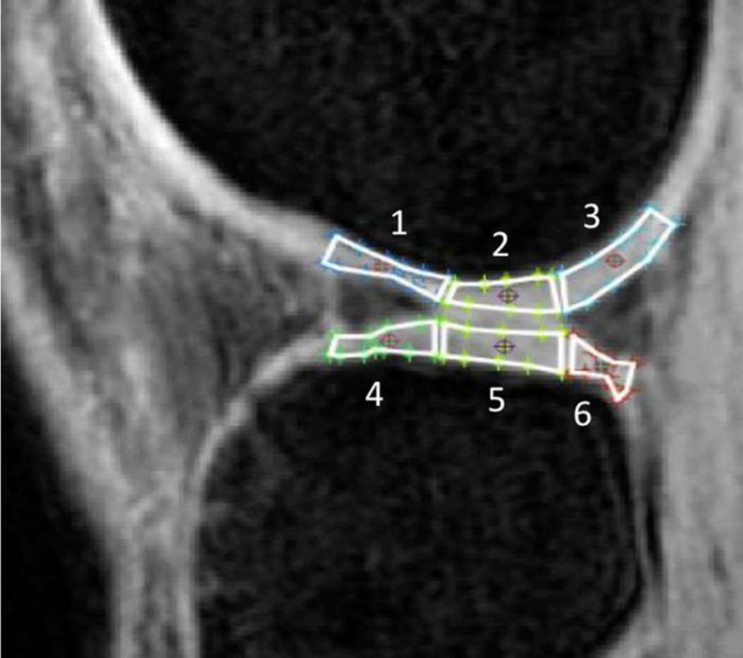
Figure 1. Representative 3D fast spin-echo image showing subcompartment definition for the lateral femoral condyle and tibia plateau.

The anterior regions (1, 4) are regions above and below the anterior horn of the meniscus, respectively; the posterior regions (3, 6) are regions above and below the posterior horn of the meniscus, respectively; the middle regions (2, 5) are between the anterior and posterior horns of the meniscus, respectively. 1, anterior femoral condyle; 2, middle femoral condyle; 3, posterior femoral condyle; 4, anterior tibial plateau; 5, middle tibial plateau; 6, posterior tibial plateau. Image with permission.29
