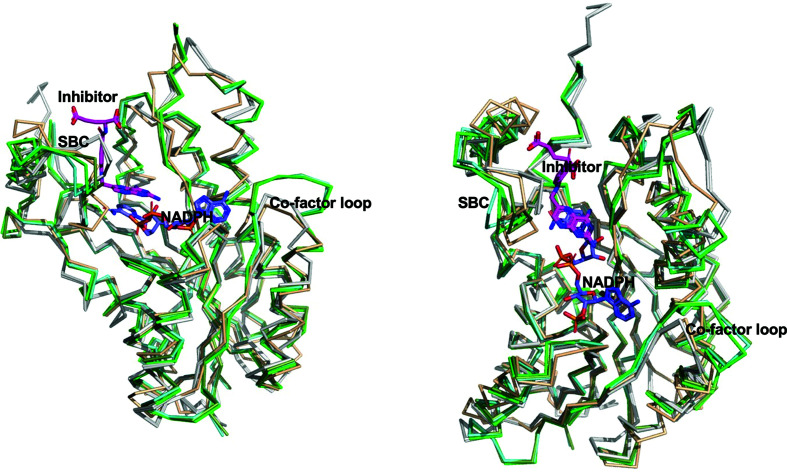
Figure 3.
Two views comparing BsFolM and BcFolM monomers with similar structures. The BsFolM and BcFolM monomers (gray) have the prototypical double-Rossmann fold of NADPH-dependent short-chain dehydrogenase/reductases observed in the molecular-replacement search model (tan) and protozoan pteridine reductase (green). The superposed protozoan structures are Trypanosoma brucei pteridine reductase with cyromazine (PDB entry 2x9n; cyan green), T. brucei pteridine reductase ternary complex with cofactor and inhibitor (PDB entry 4cm8; dark green) and T. cruzi pteridine reductase (PDB entry 1mxf; light green). The cofactor NADPH is shown in blue sticks, while the inhibitor from PDB entry 1mxf is shown as magenta sticks in the substrate-binding cavity. As in Fig. 2 ▸, SBC stands for substrate-binding cavity.

