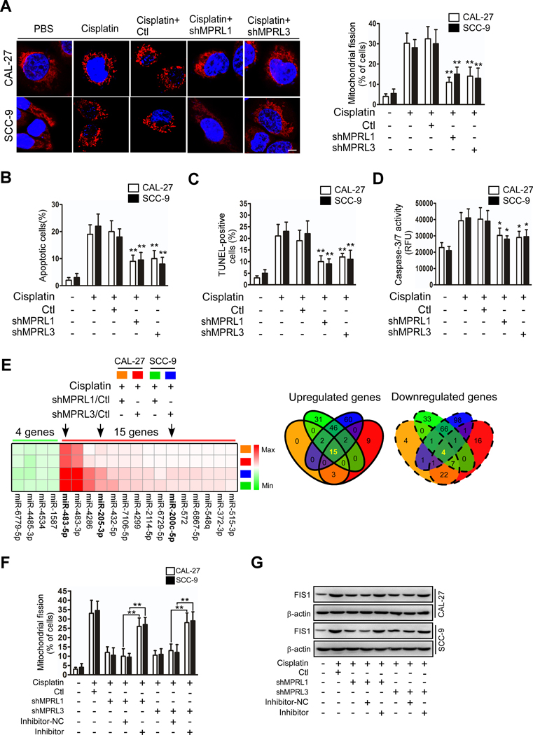
Figure 2. MPRL promotes mitochondrial fission and cisplatin sensitivity in TSCC through the miR-483–5p-FIS1 axis.
(A-D) Knockdown of MPRL attenuated mitochondrial fission and apoptosis in CAL-27 and SCC-9 cells. Mitochondrial fission was detected by staining with MitoTracker Red (left panel) and quantified (right) (A); Scale bar, 3 μm; cell apoptosis was detected using flow cytometry (B), TUNEL (C), and caspase-3/7 activity assays (D). (E) Target miRNAs of MPRL were screened by microarray in cells treated with cisplatin. Heat map (left panel) and Venn diagrams (right) depicted differentially expressed miRNAs in cisplatin-treated CAL-27 and SCC-9 cells stably expressing shMPRLs (fold change ≥1.5). (F) The inhibitory effect of MPRL knockdown on mitochondrial fission was attenuated after inhibiting miR-483–5p levels. Mitochondrial fission was detected by staining with MitoTracker Red. (G) Western blot analysis showed that the inhibitory effect of MPRL knockdown on FIS1 expression was attenuated after inhibiting miR-483–5p levels. *P<0.01 and **P<0.001, 2-tailed Student’s t tests.

