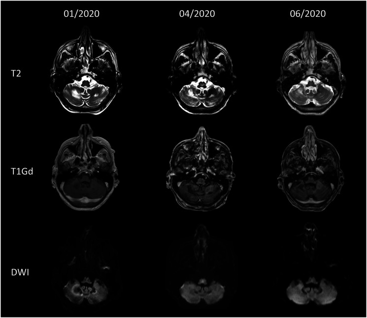
Figure 1.
Magnetic resonance imaging findings over time. Upper row: T2-weighted axial images show hyperintense white-matter lesions in the cerebellum. Middle row: Axial T1 gadolinium (Gd)-weighted images show no Gd-enhancement. Lower row: Axial diffusion weighted image shows no restricted diffusion.

