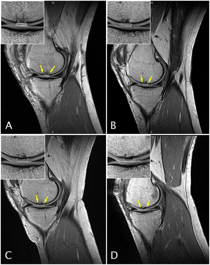
Figure 3.
Sagittal magnetic resonance (MR) images obtained with a proton density weighted (PDw) fast spin echo (FSE) sequence of a 51-year-old female patient at study entry with a chondral defect of 1 cm2 on the medial femoral condyle who underwent cartilage repair surgery with GelrinC. MR imaging was performed at baseline (A), 6 (B), 12 (C), and 24 (D) months after surgery.

