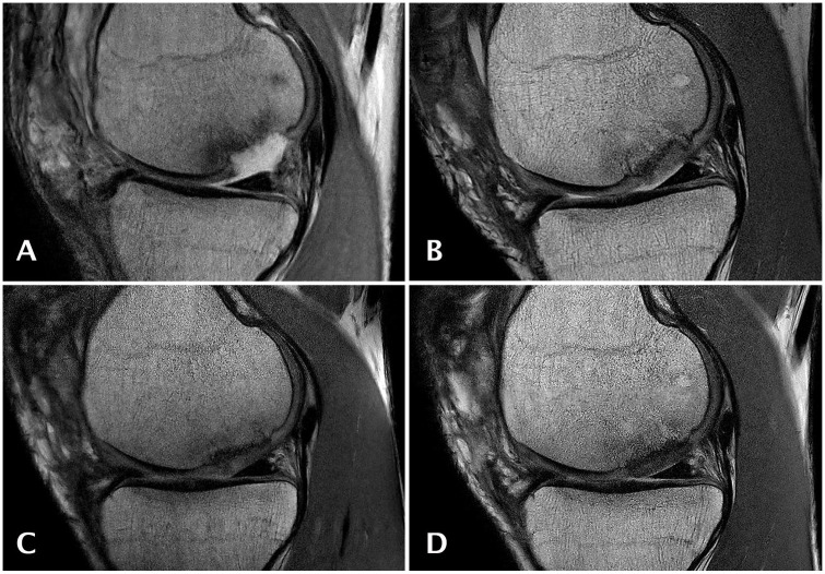
Figure 4.
Sagittal magnetic resonance (MR) images obtained with a proton density weighted (PDw) fast spin echo (FSE) sequence of an 18-year-old male patient at study entry with an osteochondral lesion of 2.8 cm2 on the medial femoral condyle who underwent cartilage repair surgery using GelrinC. MR imaging was performed at baseline (A), 6 (B), 12 (C), and 24 (D) months after surgery.

