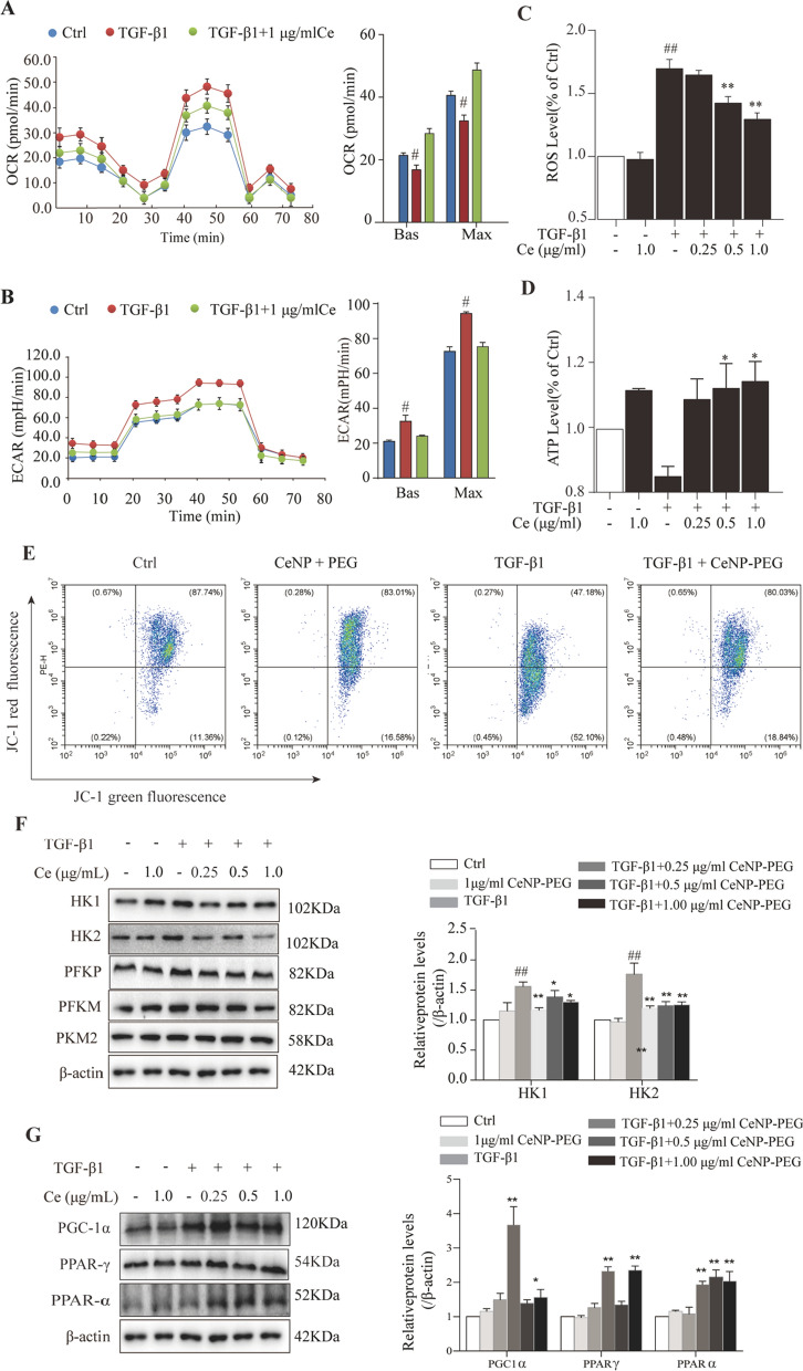
Fig. 4.
CeNP-PEG blocked the aerobic glycolysis and enhances OXPHOS, contributing to EMT suppression in vitro A The OCR measurements of the mitochondrial stress test and B The ECAR measurements of the glycolysis stress test were performed in HK-2 cells treated with or without TGF-β1, and/or CeNP-PEG. The basic and maximum capacity of OCR and ECAR were quantified. C The ROS levels of cells treated with TGF-β1 and/or CeNP-PEG was determined using the fluorescence spectrophotometry. D The ATP content of cells treated with TGF-β1 and/or CeNP-PEG was determined using the ATP Bioluminescence Assay Kit. E Mitochondrial membrane potential of cells treated with TGF-β1 and/or CeNP-PEG was measured by flow cytometry. F and G The expression of HK1/2, PFKP, PFKM and PKM2, total-Samd2/3 and p-Samd2/3 stimulated by TGF-β1 and/or CeNP-PEG was evaluated by western blot analysis. β-actin was used as the loading control, and all data were presented as mean ± SD (#P < 0.05 and ##P < 0.01 for TGF-β1 vs. control, and *P < 0.05 and **P < 0.01 for TGF-β1 + CeNP-PEG; n = 3 for each group)

